

Filter articles
标签
产品
Loading...
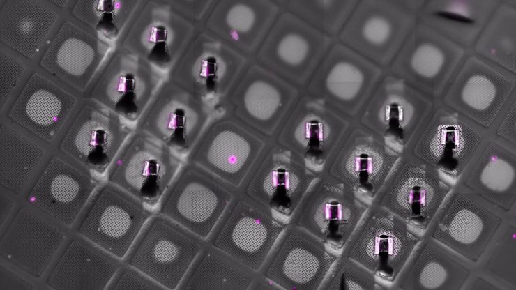
Fluorescence-Guided Cryo-ET of Malaria Parasites
Correlative cryo workflow with cryo-planing and cryo-fluorescence imaging enables targeted cryo-ET of parasite-rich regions in complex tissue.
Loading...
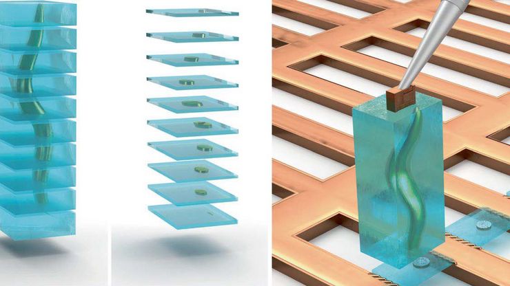
Cryo-ET Sample Preparation: From Waffle Method to Serial Lift-Out
Cryo-ET sample preparation becomes more demanding when specimens are thicker, larger, or more complex. This webinar brings together four perspectives on how high-pressure freezing can be connected…
Loading...

Waffle Method Workflow: From HPF to Cryo-ET Lamellae
Waffle freezing provides an HPF-based route to cryo-ET sample preparation. This workflow guide follows the process from grid and carrier assembly to vitrification, cryo-FIB milling, lamella…
Loading...
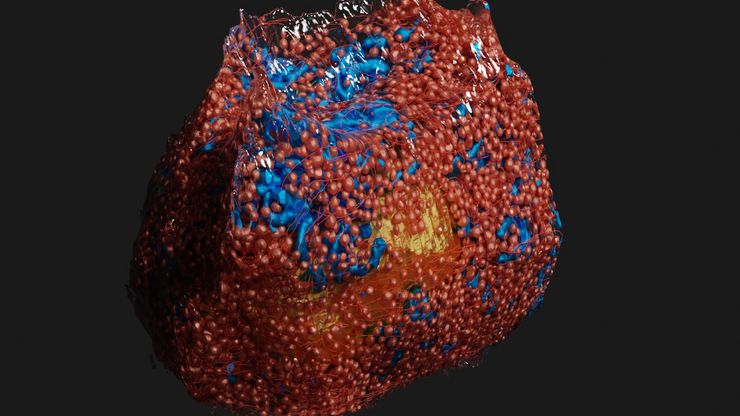
High-Pressure Freezing Protocols for Ultrastructural 3D EM
High pressure freezing (HPF) can help preserve hydrated cells and tissues close to their biological state at the moment of immobilization, supporting more reliable ultrastructural interpretation than…
Loading...
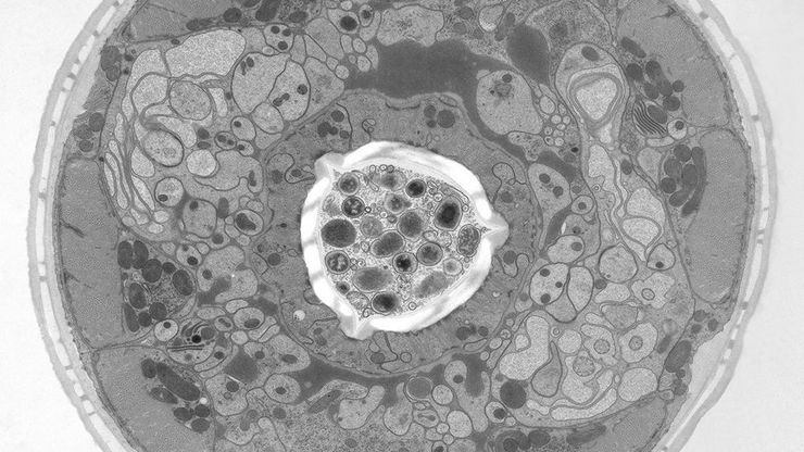
高压冷冻简介
水是细胞最主要的组成部分,因此对于维持细胞超微结构至关重要。目前,冷冻固定是固定细胞成分,而不导致其显著结构变化的唯一途径。现阶段有两种常见的方法:投入冷冻与高压冷冻固定。
Loading...
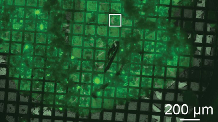
"Waffle方法":使用高压冷冻制备复杂样品
本文介绍了一种特殊的高压冷冻方法,即 "Waffle 方法 "的优点。了解 "Waffle 方法 "如何使用电镜载网作为高压冷冻的载体,从而减少样本厚度并支持复杂生物样本的高效低温电子显微镜工作流程。此外,本文还强调了现代 HPF 系统-徕卡微系统公司 EM ICE 的优势,并列举了 EM ICE 用于 "Waffle方法 "的参考文献。
Loading...
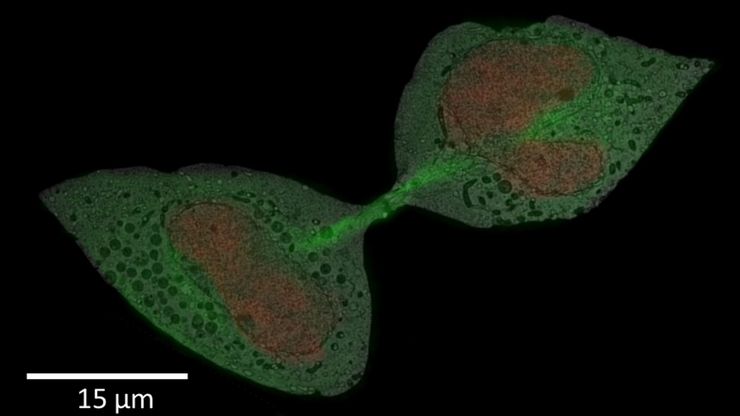
如何成功进行活细胞光电关联
Coral Life 提供了简化的活细胞 CLEM 解决方案,用于深入了解细胞成分随时间发生的结构变化。除了工作流程手册中描述的技术处理外,本文还提供了成功进行实验的其他知识。
Loading...

如何使用Coral Life(活细胞光电联用)改进活细胞成像
对于活细胞 CLEM 应用而言,光学显微镜成像是在正确的时间以正确的状态识别正确细胞的关键步骤。在本文中,徕卡专家就使用宽场系统的优势以及使用蓝宝石作为细胞培养基底时需要克服的障碍分享了他们的见解。
Loading...
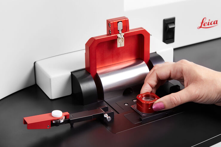
如何让样品保持在生理状态
Coral Life工作流将动态数据与最佳的样品固定方式(高压冷冻)相结合。然而,如果您的细胞因为温度下降,或缺氧气、二氧化碳或营养物质缺乏而受到损伤,那么再好的样品保存也没有意义。这些因素将影响一系列的生物过程,甚至破坏原超微结构基础,影响您的分析。


