
联系我们
联系当地专家,获取有关符合您需求和预算的专家建议

,有丝分裂纺锤体(黄色,EGFP),高尔基体(红色,Atto647N),线粒体(绿色,AF532),肌动蛋白丝(紫色,SiR700)。")
Featured image
C2C12 细胞成像
细胞使用核纤层蛋白 B(紫色)、Hoechst(蓝色)和 γH2AX(黄色)染色,分别表示细胞核结构、DNA 和 DNA 损伤。 细胞使用 THUNDER 活细胞成像系统和 63 倍/1.4 数值孔径的油镜成像。 图像由加州大学戴维斯分校生物科学学院神经生物学、生理学与行为学系 Lucas Smith 博士提供。
Follow us on Instagram
相关文章
癌症研究中显微镜的历史、发展和趋势
癌症是一种全球性疾病,2020 年全球将新增 1800 万确诊病例,1000 万人死于癌症。预计到 2040 年,病例数将增加约 55%。研究了解癌症的发生、发展以及开发新的诊断工具和治疗方法至关重要。
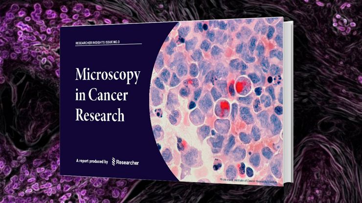
Researchers Insights: Microscopy in Cancer Research
Discover how imaging techniques are driving cancer research forward. In this issue, we present comprehensive multimodal studies using microscopy, as well as new directions in intraoperative cancer…

人工智能与深度视觉蛋白质组学 (DVP) 相结合,推进疾病研究
在这次网络研讨会上,Andreas Mund 博士将介绍深度可视蛋白质组学(DVP)--一种将人工智能驱动的组织空间分辨、非靶向蛋白质组学相结合的尖端平台。他展示了 DVP 如何从最小的、表型匹配的细胞群中识别数千种蛋白质,并在复杂的临床组织样本中生成高分辨率分子图谱,从而在细胞水平上解码疾病机制。
 and co-stained for nuclear DNA (Hoechst 33342), microtubules (Alexa 555) and F-actin (ATTO 643). Image was captured on Mateo FL.")
用于二维细胞培养的显微镜和AI解决方案
这本电子书探讨了显微镜和AI技术在二维细胞培养工作流程中的整合。报告重点介绍了明视野、相衬和荧光等传统成像方法如何支持常规细胞监测,而 Mateo TL 和 Mateo FL 数字式倒置显微镜则通过自动汇合检查、细胞计数和转染分析提高了可重复性。它还展示了综合数据管理、审计跟踪和样本跟踪如何改进文档和研究的完整性。本书最后展望了未来趋势,包括微流控技术和 2D-3D…
.")
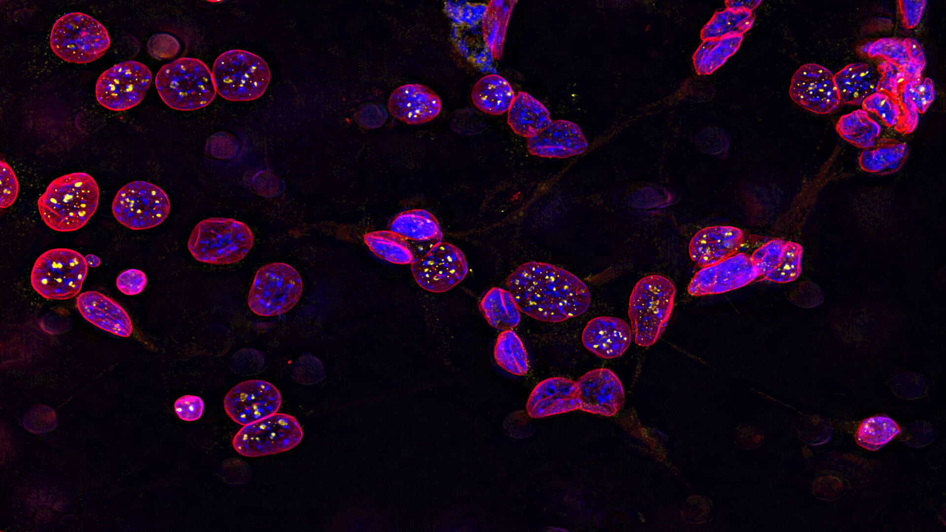
人工智能驱动的乳腺癌研究多重染色成像空间分析工具
乳腺癌(BC)是女性因癌症死亡的主要原因,研究查肿瘤微环境(TME)对于阐明肿瘤进展机制至关重要。利用超多标染色空间蛋白质组学技术系统地绘制肿瘤微环境图谱可以提高精准免疫肿瘤学的能力。在这里,我们将基于人工智能的高倍空间分析应用于BC组织,研究免疫细胞类型和生物标记物,从而深入了解受免疫疗法反应的TME分子机制。
.")
利用光片显微技术聚焦三维长时程成像
长时程三维成像揭示了复杂的多细胞系统是如何生长和发育的,以及细胞是如何随着时间的推移而移动和相互作用的,从而揭示了发育、疾病和再生方面的重要知识。光片显微镜一次只照射样品的一个薄片,大大减少了光损伤,保护了样品的活性。这种温和的高速技术可在数小时甚至数天内提供清晰的体数据,使研究人员能够实时捕捉生物学的发展过程。

体电子显微学与人工智能图像分析
该文章详细阐述了利用体电子显微镜技术 (volume-SEM) 结合人工智能辅助图像分析,对生物组织进行三维研究的工作流程。研究的重点是一种名为毛滴虫的原生动物,这是一种有鞭毛的寄生虫,是导致性传播感染——滴虫病的病原体。为了可视化其复杂的内部结构,研究人员采用了体电子显微镜技术,通过对一系列超薄切片进行成像来重建三维模型。
 at Leica Microsystems.")
空间蛋白质组学的突破如何拯救生命
中毒性表皮坏死溶解症(TEN)是一种罕见的、但对抗生素或痛风治疗等常见药物的破坏性反应。这种疾病开始时并无大碍,通常只是皮疹,但会迅速升级为大面积皮肤脱落,类似于严重烧伤。尽管 TEN病情十分严重,但其基本机制仍然难以捉摸,治疗方案也仅限于支持性护理。TEN 的死亡率高达 30%,长期以来一直是临床医生的噩梦,直到现在才有了靶向疗法。

来捕捉发育动态的3D成像
本应用说明展示了研究人员如何成功利用 Viventis Deep 双视角光片显微镜探索3D多细胞模型(包括有机体、球形体和胚胎)的高分辨率长期成像,从而为发育生物学和疾病研究带来新的可能性。
 at 2 weeks. Image acquired using Mica.")
如何为深层肌肉组织中的轴突再生成像
这项研究重点介绍了亚伦-李(Aaron Lee)博士对截肢后肌肉移植中神经再生的定位研究。肢体缺失通常会导致生活质量下降,这不仅是因为组织缺失,还因为轴突再生紊乱引起的神经性疼痛。Mica组织学成像和荧光成像可帮助了解神经再生过程中轴突的生长和分支这项研究有助于塑造未来的神经假体接口设计,改善患者的治疗效果。
