
Filter articles
主题和标签
产品
Loading...
斑马鱼研究
为了在筛选、分拣、操作和成像过程中获取高质量结果,您需要观察细节和结构,从而为您的下一步研究做出正确的决策。
徕卡体视显微镜和透射光底座以出众的光学器件和优良的分辨率而闻名,是全世界研究学者的首选。
Loading...
")
How to Streamline High-Plex Imaging for 3D Spatial Omics Advances
In this webinar, Dr. Julia Roberti and Dr. Luis Alvarez from Leica Microsystems introduce SpectraPlex, a new functionality integrated into the STELLARIS confocal platform for high-plex 3D spatial…
Loading...

Transforming Research with Spatial Proteomics Workflows
Spatial Proteomics, Nature Methods 2024 Method of the Year, is driving research advancements in cancer, immunology, and beyond. By combining positional data with high throughput imaging of proteins in…
Loading...
.")
How Fluorescence Guides Sectioning of Resin-embedded EM Samples
Electron microscopes, including transmission electron microscopes (TEM) and scanning electron microscopes (SEM), are widely utilized to gain detailed structural information about biological samples or…
Loading...

Coherent Raman Scattering Microscopy Publication List
CRS (Coherent Raman Scattering) microscopy is an umbrella term for label-free methods that image biological structures by exploiting the characteristic, intrinsic vibrational contrast of their…


Loading...
 applied. Image courtesy of Samuel East, Uncommon Bio.")
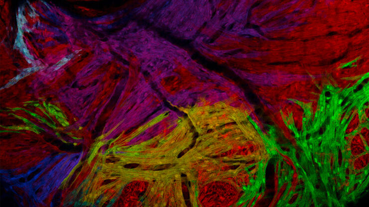
利用新型可扩展的干细胞培养设计未来
具有远见卓识的生物技术初创企业 Uncommon Bio 正在应对世界上最大的健康挑战之一:食品可持续性。在这次网络研讨会上,干细胞科学家塞缪尔-伊斯特(Samuel East)将展示他们如何使细胞农业的干细胞培养基既安全又经济可行。了解他们如何将培养基成本降低 1000 倍,并开发出不含动物成分、食品安全的 iPSC 培养基。
Loading...
, microglia (TMEM119, IBA1), and Alzheimer’s-associated markers (β-amyloid and p-Tau217).")
利用大数据探索阿尔茨海默病的空间蛋白组
阿尔茨海默病是一种遗传性和散发性的神经退行性疾病,导致中晚年认知能力下降,特征为β-淀粉样蛋白斑块和 tau蛋白 缠结。由于治疗选择有限,新的研究策略至关重要。Cell DIVE 多重成像解决方案可以对阿尔茨海默病脑组织进行研究,揭示,可能新的研究方向。这里我们展示了 Cell DIVE 多重成像仪的图像查看器,用户能够直接在自己的浏览器中访问完整的阿尔茨海默病多重数据集。
