
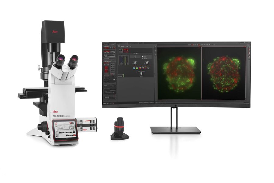
类器官和3D细胞培养
生命科学研究中最令人振奋的最新进展之一是3D细胞培养系统的发展,例如类器官、球状体或器官芯片模型。 3D细胞培养物是一种人工环境,在这种环境中,细胞能够在三维空间中生长并与周围环境相互作用。 这些环境条件与它们在体内的情况相似。 类器官是一种3D细胞培养物,包含器官特异性细胞类型,可以表现出器官的空间组织和复制器官的某些功能。 类器官重现了一个生理上高度相关的系统,使研究人员能够研究复杂的多维度问题,例如疾病发作、组织再生和器官之间的相互作用。 光学显微镜是用类器官研究复杂的相互作用与关系的重要方法。
徕卡成像解决方案支持这些多功能样本的研究,使用这些系统可进行深度快速成像,适合终点测量或者通过活细胞成像进行动力学研究。
联系我们
联系当地专家,获取有关符合您需求和预算的专家建议
克服类器官成像中的挑战
成像是研究类器官和球状体等3D细胞培养物的关键技术。
类器官的有效成像构成一系列的新挑战,因为它们包含很大的体积。 类器官可以被固定、进行免疫标记,并使用透明技术进行研究,以便对其3D结构成像。 通常,这些研究使用共聚焦显微镜进行,因为对于宽场系统而言,对细胞层多于2-3个的培养物成像可能具有很大的挑战性,宽场系统固有的模糊现象会掩盖感兴趣的信号。
类器官也可以用来研究动态过程。 活体类器官研究会面临典型的成像问题,例如光毒性和低信噪比,特别是在样本深度成像时。 最近,各种快速采集显微成像方法(如 FLIM 或光片)在活体类器官研究中受到青睐,因为使用这些方法时可以不改变样本的生理机能。
.提供")
相关文章
History, Developments and Trends of Microscopy in Cancer Research
Cancer is a global disease, with 18 million new cases diagnosed and 10 million cancer-related deaths worldwide in 2020. This burden is set to increase, with a projected increase in cases of ~55% by…
, tubulin with Cy5 (red), and nuclei with DAPI (blue). Image courtesy of Dr. Günter Giese, Max Planck Institute for Medical Research, Heidelberg, Germany.")
荧光染料应用和特性概述
本文将介绍常用的荧光染料并概述其特性。荧光显微镜借助荧光染料、荧光蛋白或使用抗体的免疫荧光染色来研究特定的细胞成分。由于荧光剂种类繁多,荧光显微镜可用于检查蛋白质、核酸、聚糖、细胞器和其他细胞结构。
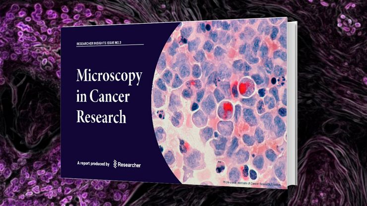
Researchers Insights: Microscopy in Cancer Research
Discover how imaging techniques are driving cancer research forward. In this issue, we present comprehensive multimodal studies using microscopy, as well as new directions in intraoperative cancer…
 image of a cross section of C. elegans (roundworm). Courtesy of T. Müller-Reichert, MPI-CBG, Dresden, Germany and K. McDonald, University of California, Berkeley, USA.")
高压冷冻简介
水是细胞最主要的组成部分,因此对于维持细胞超微结构至关重要。目前,冷冻固定是固定细胞成分,而不导致其显著结构变化的唯一途径。现阶段有两种常见的方法:投入冷冻与高压冷冻固定。
.")
利用光片显微技术聚焦三维长时程成像
长时程三维成像揭示了复杂的多细胞系统是如何生长和发育的,以及细胞是如何随着时间的推移而移动和相互作用的,从而揭示了发育、疾病和再生方面的重要知识。光片显微镜一次只照射样品的一个薄片,大大减少了光损伤,保护了样品的活性。这种温和的高速技术可在数小时甚至数天内提供清晰的体数据,使研究人员能够实时捕捉生物学的发展过程。
 and tubulin (magenta), acquired using Viventis Deep. Courtesy of Akanksha Jain, Treutlein Lab ETH-DBSSE Basel (Switzerland).")
如何深入了解类器官和细胞球模型
在本电子书中,您将了解3D细胞培养模型(如类器官和细胞球)成像的关键注意事项。探索创新型显微镜解决方案,来实时记录类器官和细胞球的动态成像过程。

来捕捉发育动态的3D成像
本应用说明展示了研究人员如何成功利用 Viventis Deep 双视角光片显微镜探索3D多细胞模型(包括有机体、球形体和胚胎)的高分辨率长期成像,从而为发育生物学和疾病研究带来新的可能性。
 at 2 weeks. Image acquired using Mica.")
如何为深层肌肉组织中的轴突再生成像
这项研究重点介绍了亚伦-李(Aaron Lee)博士对截肢后肌肉移植中神经再生的定位研究。肢体缺失通常会导致生活质量下降,这不仅是因为组织缺失,还因为轴突再生紊乱引起的神经性疼痛。Mica组织学成像和荧光成像可帮助了解神经再生过程中轴突的生长和分支这项研究有助于塑造未来的神经假体接口设计,改善患者的治疗效果。

用于三维生物成像的集成连续切片与冷冻电镜工作流程
本场网络研讨会探讨了集成化工具如何支持从样品制备到图像分析的电子显微镜全流程。专家Andreia Pinto博士、Adrian Boey博士与Hoyin Lai博士将介绍UC Enuity超薄切片机和Aivia图像分析平台,并演示这些工具如何同时适用于常温与低温实验环境。会议内容包含阵列断层成像、基于深度学习的图像分割、以及生物成像中cryo-lift-out工作流程的实际案例解析。

利用人工智能图像分析工具更快、更轻松地获得洞察力
了解 Aivia 如何通过快速设置、准确的人工智能检测和简便的批量处理功能,帮助科学家简化图像分析。

揭开类器官模型在生物医学研究中的秘密
准备深入了解类器官和3D培养物的世界,它们是促进我们了解人类健康的重要工具。浏览这些复杂的结构并获取清晰的图像进行分析是一项挑战。在本次活动中,来自牛津大学和伦敦大学学院的研究人员将与我们一起展示Thunder Imager Cell转盘共聚焦系统 如何提供更有说服力的高质量数据,以便深入了解各种模型。
 applied. Image courtesy of Samuel East, Uncommon Bio.")
利用新型可扩展的干细胞培养设计未来
具有远见卓识的生物技术初创企业 Uncommon Bio 正在应对世界上最大的健康挑战之一:食品可持续性。在这次网络研讨会上,干细胞科学家塞缪尔-伊斯特(Samuel East)将展示他们如何使细胞农业的干细胞培养基既安全又经济可行。了解他们如何将培养基成本降低 1000 倍,并开发出不含动物成分、食品安全的 iPSC 培养基。

Mica: 助力伦敦帝国学院开展跨学科科研研究
这篇访谈重点介绍了伦敦帝国学院的 Mica 所产生的变革性影响。科学家们解释了Mica如何改变了游戏规则,扩大了研究的可能性,促进了跨学科合作。他们解释了使用 Mica 进行详细的活细胞成像如何提供更有意义的信息,使科学家始终站在研究的最前沿。研究小组预计,Mica将继续开辟新的研究途径,包括研究微流体技术和其他先进应用。
")
利用子宫内膜类器官推进子宫再生疗法
康教授团队致力于研究决定子宫微环境的关键因素,该环境对胚胎着床和妊娠维持至关重要。他们正为罹患阿什曼综合征等子宫内膜疾病的患者开发恢复子宫内膜功能的新型治疗策略。通过将 3D 子宫内膜类器官移植至小鼠模型,该团队揭示了子宫内膜强大的再生能力的细胞与分子机制。本次访谈将深入探讨其团队的研究内容及Mica在研究中所发挥的重要作用。
.")
您的 3D 类器官成像和分析工作流程效率如何?
类器官模型已经改变了生命科学研究,但优化图像分析协议仍然是一个关键挑战。本次网络研讨会探讨了类器官研究的简化工作流程,首先是实时的三维细胞培养检查,接下来是高速、高分辨率的三维成像,生成清晰的图像和更纯净的数据,以便对生长速率、细胞迁移和三维细胞相互作用等参数进行准确地人工智能分割和量化,从而实现更深入的洞察。

在神经发育过程中,细胞是如何相互交流的?
细胞间通信是大脑发育过程中一个必不可少的过程,它受到多种因素的影响,包括细胞的形态、粘附分子、局部细胞外基质和分泌囊泡。在本次网络研讨会上,您将了解到对这些机制更深入的理解是如何推动对神经发育障碍的理解的。
