Gold EM grid with a silicon dioxide (SiO₂) support film, containing vitrified T47D cells intrinsically expressing GFP-H2B (labeling nuclei) and RFP-LifeAct (labeling the actin cytoskeleton) and high-pressure frozen C. elegans germline tissue, fluorescently labeled with GFP-tagged synaptonemal complex protein

电子显微镜样品制备工作流程& 用途

通过使用徕卡样品制备解决方案,研究人员可以在利用电子显微镜对样品进行成像时,始终获得高质量、精确且可重复的结果。我们的解决方案支持扫描电子显微镜(SEM)、透射电子显微镜(TEM)及冷冻电镜(Cryo EM)。

请联系我们!

我们经验丰富的成像专家团队,竭诚为您提供关于电镜样品制备工作流程与应用的解决方案咨询。
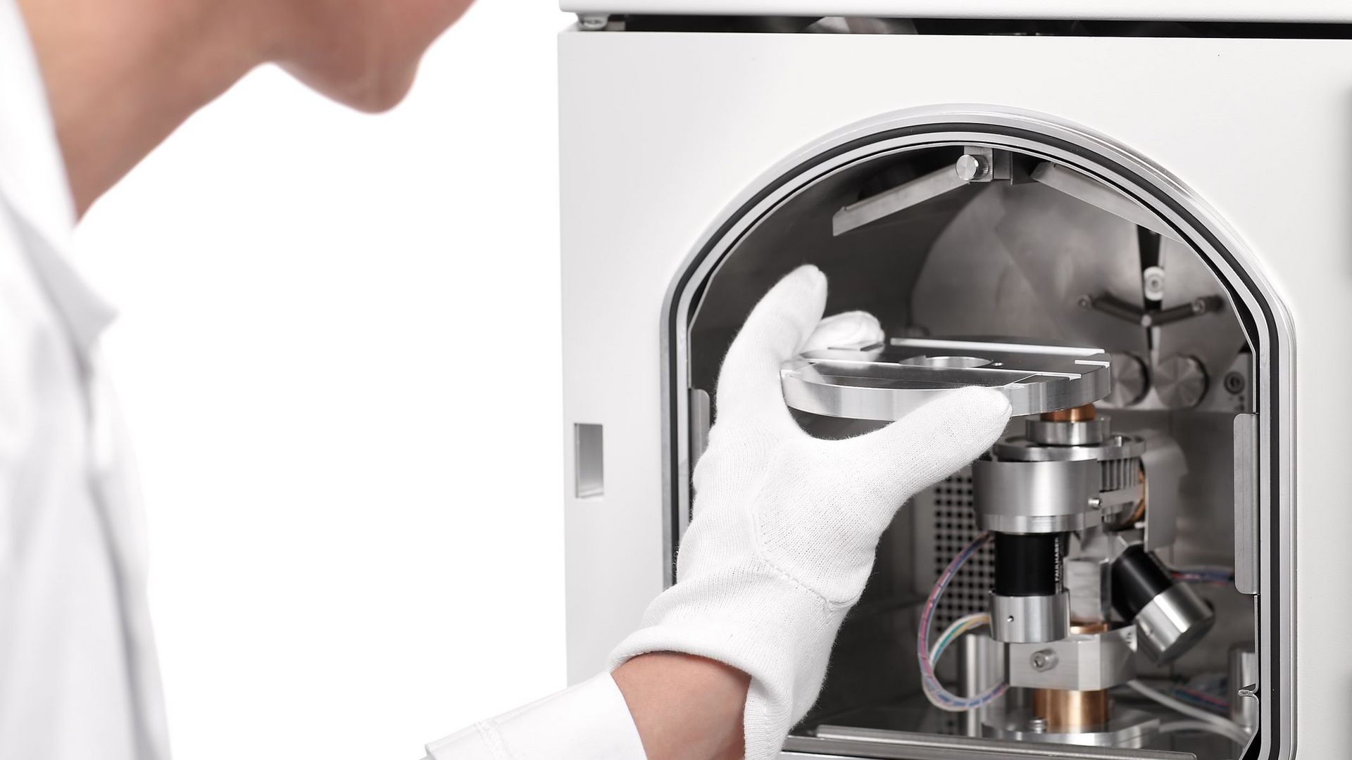
研究人员如何制备用于扫描电镜的样品?

扫描电子显微镜(SEM)的样品制备涉及镀膜、干燥及包埋等技术,以确保获得最佳成像效果。通过使用徕卡解决方案制备扫描电镜样品,研究人员可以提高样品导电性、减少伪影,并确保获得稳定、高质量的样品表面,从而在常温下实现可靠的SEM成像。

透射电镜(TEM) 样品制备的最佳方法是什么?

TEM(透射电子显微镜)样品制备需要制备超薄、高度无损的切片,对电镜载网进行溅射镀膜,并结合染色技术,以实现对样品精细结构的清晰可视化。凭借徕卡仪器的卓越精准度与高度可靠性,研究人员能够获取高分辨率TEM图像。
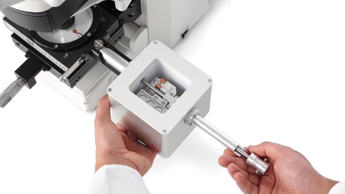
冷冻电镜样品制备的关键是什么?

冷冻电镜通过快速冷冻实现玻璃化,使样品结构得以保持其天然状态。

徕卡冷冻制备解决方案——涵盖先进的玻璃化、镀膜、超薄切片、冷冻平面加工,以及可定制的冷冻传输和CLEM工作流程——确保获得可重复、无污染的样品。这些解决方案能够帮助保持样品的天然结构,实现精准定位,并支持高分辨率冷冻电镜成像。

为什么选择徕卡仪器及解决方案进行电镜样品制备?


扫描电镜(SEM)样品制备

徕卡显微系统提供的SEM制备工具包括高真空溅射镀膜仪和临界点干燥仪。这些解决方案确保获得高质量的表面成像效果。此外,徕卡仪器能为您的SEM研究提供稳定、可靠的结果。


透射电镜(TEM)样品制备

研究人员可以信赖徕卡显微系统的TEM制备解决方案,以获得精确与可靠的结果。超薄切片机和自动染色系统等核心工具,能够高效制备超薄切片,助力获取高分辨率图像。溅射镀膜可提高样品导电性,从而提升成像质量。


全方位冷冻电镜(Cryo-EM)样品制备

通过使用徕卡显微系统的冷冻制备仪器——包括投入式冷冻仪和高压冷冻仪——研究人员能够实现样品的快速冷冻与玻璃化。此外,我们的冷冻工作流程样品传输系统为各类样品提供多样化的传输方案。这一传输方案可确保样品免受污染,为精确、细致的Cryo-EM分析提供可靠保障。

相关文章

查看我们关于电镜样品制备工作流程与应用的最新文章

徕卡显微系统的知识门户网站提供有关显微镜学的科学研究资料和教学材料。网站内容专门面向初学者、经验丰富的从业者和科学家,为他们的日常工作和实验提供支持。

更多文章
3D rendering of a mouse beta cell with mitochondria (blue)
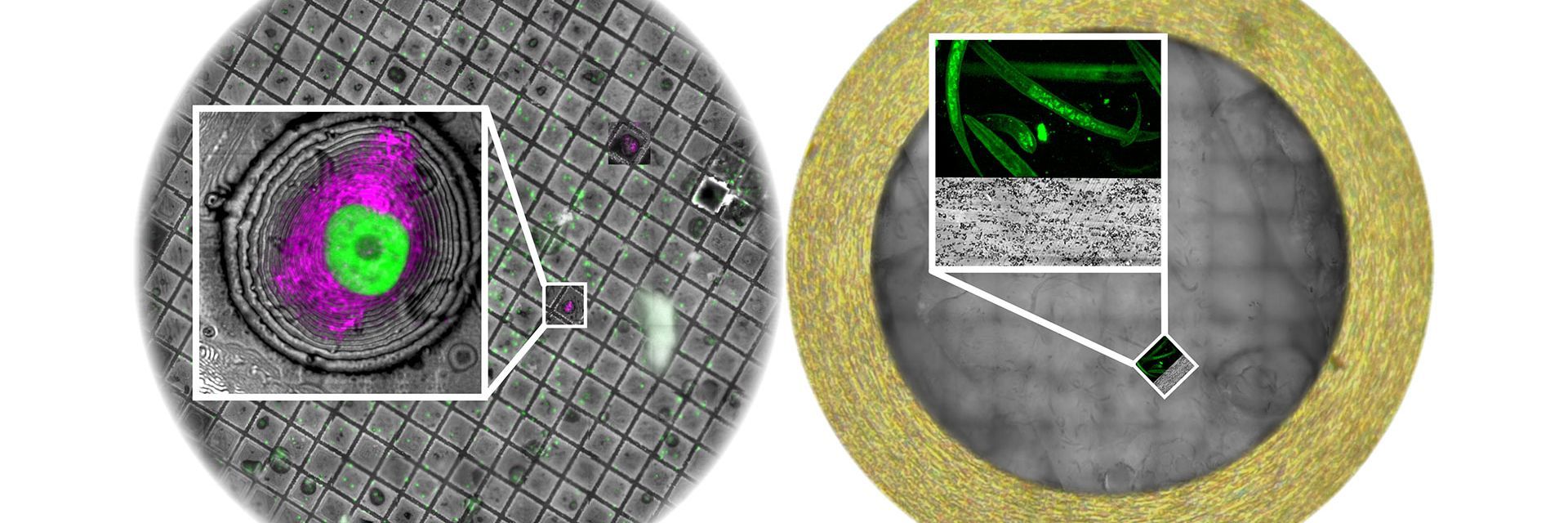
, insulin SGs (orange), microtubules (red), nucleus (yellow), and plasma membrane (transparent).

High-Pressure Freezing Protocols for Ultrastructural 3D EM

High pressure freezing (HPF) can help preserve hydrated cells and tissues close to their biological state at the moment of immobilization, supporting more reliable ultrastructural interpretation than…
Claudia Mayrhofer ZFE Graz

Ultramicrotome UC Enuity in Practice: Stable 15 nm Sections at ZFE

After using the UCT and UC6 ultramicrotomes, Claudia Mayrhofer calls UC Enuity a leap in stability—so robust that vibrations and temperature shifts don’t spoil sections, even with multiple users. Auto…
Sectioning resin-embedded sambles

超薄切片技术电子书:定位、修块& 对刀

超薄切片技术正经历日新月异的发展,当今的显微镜系统对高质量切片、精准定位以及可重复的工作流程提出了更高要求。这本电子书整合了专家应用指南、自动化方法及实操指导,旨在帮助从初学者到资深镜检人员的每一位用户,在电镜、光电联用及体电镜工作流程中,获得一致且可靠的超薄切片。
Foraminifera (Ammonia confertitesta) 
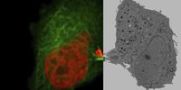
labeled with membrane-permeable calcein, high-pressure frozen in salt water using EM ICE. The sample was cryo-planed and targeted with the M205 on the Cryo-Fluo Enuity, then transferred under cryo conditions to the Cryo-Stellaris for widefield and confocal imaging, revealing details of the staining pattern. Image courtesy: David Evans, University of Southampton.

High-Pressure Freezing for Organoids: Cryo CLEM & FIB Lift Out

Master cryo EM workflow steps for challenging 3D samples: when to choose HPF vs. plunge freezing, reproducible blotting/ice control, contamination aware transfers, Cryo CLEM 3D targeting in organoids,…
Electron microscope (EM) 
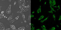
image of a cross section of C. elegans (roundworm). Courtesy of T. Müller-Reichert, MPI-CBG, Dresden, Germany and K. McDonald, University of California, Berkeley, USA.

高压冷冻简介

水是细胞最主要的组成部分,因此对于维持细胞超微结构至关重要。目前,冷冻固定是固定细胞成分,而不导致其显著结构变化的唯一途径。现阶段有两种常见的方法:投入冷冻与高压冷冻固定。
TEM micrographs of polymer sections. Left: Poly(styrene)-
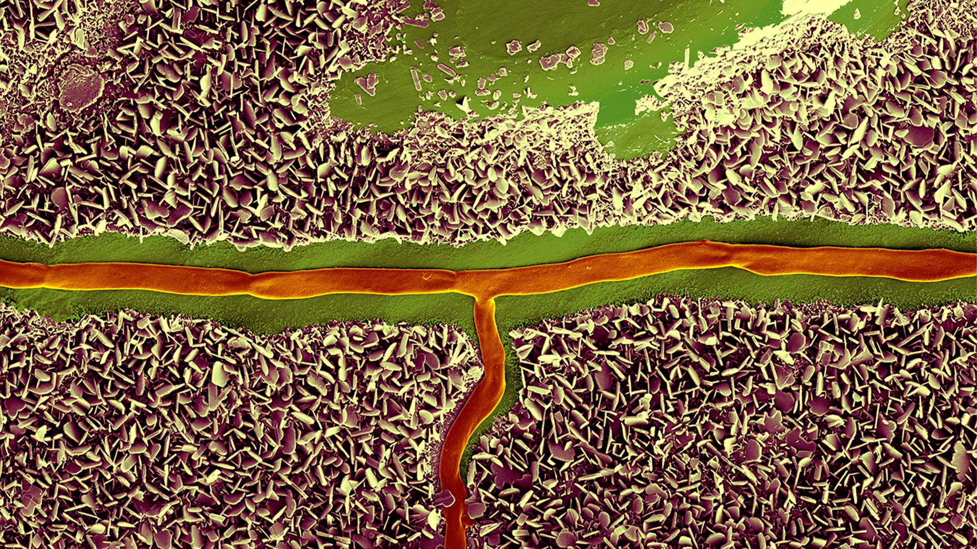
b-poly(isoprene). Right: Poly(styrene)-b-poly(methyl methacrylate).

聚合物透射电镜分析用超薄切片技术

本文全面展示了徕卡UC Enuity超薄切片机在聚合物样品超薄切片制备中的优异表现,无论是常温还是低温环境,它都能提供理想的分析样本。文中展示的高分辨率二维及三维TEM图像,有力印证了该仪器在聚合物结构分析领域,对于获得精确、可重复的样品制备结果不可或缺。
Final Segmentation of organelles in Trichomonas species. Magenta – costa, light blue – hydrogenosomes, turquoise – ER, red – vacuoles, yellow – axostyle, green – Golgi apparatus.  Sample courtesy of Isabelle Guerin-Bonne, Low Kay En, Electron Microscopy Unit, Yong Loo Lin School of Medicine, National University of Singapore. Scale bar: 1 µm.

体电子显微学与人工智能图像分析

该文章详细阐述了利用体电子显微镜技术 (volume-SEM) 结合人工智能辅助图像分析,对生物组织进行三维研究的工作流程。研究的重点是一种名为毛滴虫的原生动物,这是一种有鞭毛的寄生虫,是导致性传播感染——滴虫病的病原体。为了可视化其复杂的内部结构,研究人员采用了体电子显微镜技术,通过对一系列超薄切片进行成像来重建三维模型。

用于三维生物成像的集成连续切片与冷冻电镜工作流程

本场网络研讨会探讨了集成化工具如何支持从样品制备到图像分析的电子显微镜全流程。专家Andreia Pinto博士、Adrian Boey博士与Hoyin Lai博士将介绍UC Enuity超薄切片机和Aivia图像分析平台,并演示这些工具如何同时适用于常温与低温实验环境。会议内容包含阵列断层成像、基于深度学习的图像分割、以及生物成像中cryo-lift-out工作流程的实际案例解析。
SEM image of the full Li-NMC electrode sample, showing the two porous layers and the metal film at the center of the structure.

通过Cryo-EM(冷冻电镜)和 CryoFIB(冷冻聚焦离子束) 揭示钠电池退化机制

探索低温电镜和聚焦离子束技术如何揭示钠电池界面的内在结构。本次研讨会将提出基于隔膜渗透(而非枝晶生长)的新型退化模型,并解析电解液溶剂如何影响界面稳定性与电池性能。
使用 "Waffle方法 "进行 HPF 制备后,载网上的小鼠海马脑切片。

"Waffle方法":使用高压冷冻制备复杂样品

本文介绍了一种特殊的高压冷冻方法,即 "Waffle 方法 "的优点。了解 "Waffle 方法 "如何使用电镜载网作为高压冷冻的载体,从而减少样本厚度并支持复杂生物样本的高效低温电子显微镜工作流程。此外,本文还强调了现代 HPF 系统-徕卡微系统公司 EM ICE 的优势,并列举了 EM ICE 用于 "Waffle方法 "的参考文献。
14: 7-day old 3D cell culture of MDCK cells stably transfected with; Mx1-GFP (green), SPY555-Actin (yellow) and WGA-AlexaTM 647 (magenta), and imaged using a THUNDER Imager Cell Spinning Disk. Shown are both the spinning disk image (left) and the same image after THUNDER Computational Clearing was applied (right); scale bar 50 μm.

类器官和 3D 细胞培养

生命科学研究中最令人振奋的最新进展之一是 3D 细胞培养系统的发展,例如类器官、球状体或器官芯片模型。 3D 细胞培养物是一种人工环境,在这种环境中,细胞能够在三维空间中生长并与周围环境相互作用。 这些环境条件与它们在体内的情况相似。

了解详情

Image of C2C12 cells: The cells are stained with lamin B (magenta) which indicates nuclear structure, Hoechst (blue) indicating DNA, and γH2AX (yellow) indicating damage to DNA. Cells were imaged using a THUNDER Imager 3D Live Cell with a 63X/1.4 oil immersion objective.

细胞生物学研究

 细胞生物学成像是运用一系列的光学显微镜和电子显微镜完成的。 徕卡显微系统公司推出的成像解决方案专为扩展您的细胞生物学研究而设计。

了解详情

Mammalian cells expressing H2B-mCherry and alphaTubulin-mEGFP to visualize DNA and microtubules respectively. Left: Widefield imaging using THUNDER Imager. Right: Sample after correlated EM imaging using Leica Microsystems Coral Life workflow. Cells were cryo-immobilized by high-pressure freezing 60 min after the onset of cytokinesis. The intercellular bridge was imaged under TEM after freeze substitution and resin infiltration.

光电关联显微技术 (CLEM)

徕卡显微系统的CLEM解决方案可确保样本活性、进行质量检查,确保三维目标定位机制精确可靠。用户可以利用这些解决方案的优势,在合适的时间立即识别合适的细胞,获得高分辨率的冷冻共聚焦数据,或将荧光信息与超微结构环境结合起来。

了解详情

细胞培养

在实验室环境下培养细胞是科学家在细胞生物学、癌症研究、发育生物学等任何类型生命科学及药物研究领域开展工作的基础。了解徕卡如何帮助您在实验室中培养动物细胞。

了解详情

14: 7-day old 3D cell culture of MDCK cells stably transfected with; Mx1-GFP (green), SPY555-Actin (yellow) and WGA-AlexaTM 647 (magenta), and imaged using a THUNDER Imager Cell Spinning Disk. Shown are both the spinning disk image (left) and the same image after THUNDER Computational Clearing was applied (right); scale bar 50 μm.
Image of C2C12 cells: The cells are stained with lamin B (magenta) which indicates nuclear structure, Hoechst (blue) indicating DNA, and γH2AX (yellow) indicating damage to DNA. Cells were imaged using a THUNDER Imager 3D Live Cell with a 63X/1.4 oil immersion objective.
Mammalian cells expressing H2B-mCherry and alphaTubulin-mEGFP to visualize DNA and microtubules respectively. Left: Widefield imaging using THUNDER Imager. Right: Sample after correlated EM imaging using Leica Microsystems Coral Life workflow. Cells were cryo-immobilized by high-pressure freezing 60 min after the onset of cytokinesis. The intercellular bridge was imaged under TEM after freeze substitution and resin infiltration.
Scroll to top