

Filter articles
标签
产品
Loading...
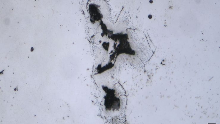
AI-Based Particle Counting with the Aivaro AI Feature Detection Software Module
The Aivaro AI Feature Detection software module enables fast manual microscope inspection of particulate contamination on various material samples. By training different classes, reflective particles,…
Loading...
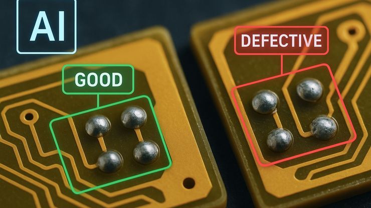
AI Image Analysis for Reliable Defect Detection
AI-based visual inspection with microscopy helps make quality control in industrial environments efficient by reducing manual inspection effort concerning defect detection, classification, and…
Loading...

An Overview of Artificial Intelligence (AI) and Machine Learning
AI is rapidly changing industrial inspection, quality control, and manufacturing workflows. But not all AI technologies work the same way. From machine learning and deep learning to the newer concept…
Loading...

Why the Future of Microscopic Inspection Is AI-Assisted, Not Fully Automated
The next wave of QC productivity gains will not come from building more automated inspection lines. Instead, it will come from augmenting millions of inspection decisions that are made manually every…
Loading...
![High-resolution 3D optics [JB19.1]with enhanced depth of field [JB20.1]allow surgeons to clearly identify and safely dissect perforators and perform delicate vessel anastomoses. Mr Dariush Nikkhah. DIEP surgery London.](/fileadmin/_processed_/8/c/csm_Bilateral_DIEP_flap_breast_reconstruction_Ghali_and_Nikkhah_9072b78814.jpg "Advanced microsurgical visualization in practice: bilateral DIEP flap breast reconstruction performed by Prof. Shadi Ghali and Mr Dariush Nikkhah.")
DIEP Flap Breast Reconstruction: Microsurgical Visualization
DIEP flap breast reconstruction is an autologous microsurgical procedure that depends on precise vessel dissection, anastomosis and perfusion assessment [1]. This article discusses how surgical…
Loading...
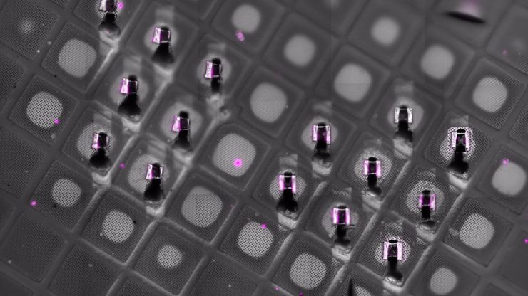
Fluorescence-Guided Cryo-ET of Malaria Parasites
Correlative cryo workflow with cryo-planing and cryo-fluorescence imaging enables targeted cryo-ET of parasite-rich regions in complex tissue.
Loading...
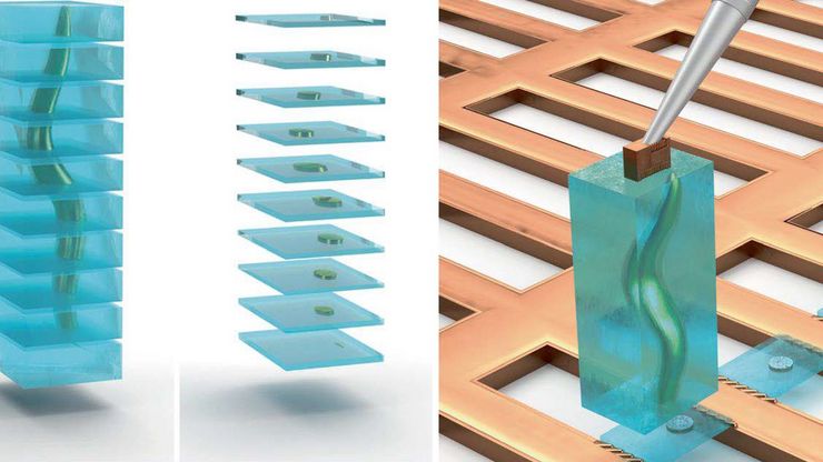
Cryo-ET Sample Preparation: From Waffle Method to Serial Lift-Out
Cryo-ET sample preparation becomes more demanding when specimens are thicker, larger, or more complex. This webinar brings together four perspectives on how high-pressure freezing can be connected…
Loading...

Waffle Method Workflow: From HPF to Cryo-ET Lamellae
Waffle freezing provides an HPF-based route to cryo-ET sample preparation. This workflow guide follows the process from grid and carrier assembly to vitrification, cryo-FIB milling, lamella…


