Filter articles
标签
产品
Loading...

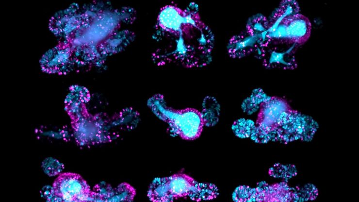
利用光片显微技术聚焦三维长时程成像
长时程三维成像揭示了复杂的多细胞系统是如何生长和发育的,以及细胞是如何随着时间的推移而移动和相互作用的,从而揭示了发育、疾病和再生方面的重要知识。光片显微镜一次只照射样品的一个薄片,大大减少了光损伤,保护了样品的活性。这种温和的高速技术可在数小时甚至数天内提供清晰的体数据,使研究人员能够实时捕捉生物学的发展过程。
Loading...

A Guide to C. elegans Research – Working with Nematodes
Efficient microscopy techniques for C. elegans research are outlined in this guide. As a widely used model organism with about 70% gene homology to humans, the nematode Caenorhabditis elegans (also…
Loading...

A Novel Laser-Based Method for Studying Optic Nerve Regeneration
Optic nerve regeneration is a major challenge in neurobiology due to the limited self-repair capacity of the mammalian central nervous system (CNS) and the inconsistency of traditional injury models.…
Loading...

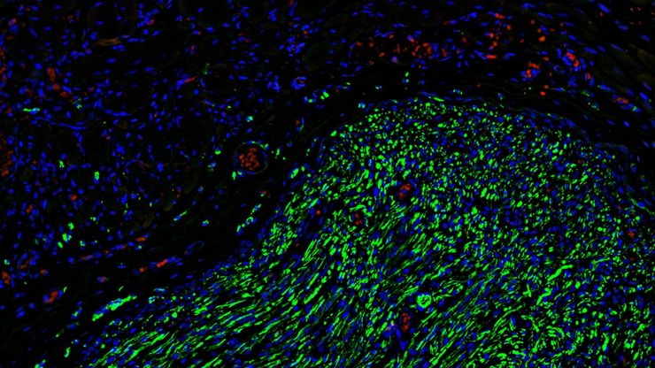
如何为深层肌肉组织中的轴突再生成像
这项研究重点介绍了亚伦-李(Aaron Lee)博士对截肢后肌肉移植中神经再生的定位研究。肢体缺失通常会导致生活质量下降,这不仅是因为组织缺失,还因为轴突再生紊乱引起的神经性疼痛。Mica组织学成像和荧光成像可帮助了解神经再生过程中轴突的生长和分支这项研究有助于塑造未来的神经假体接口设计,改善患者的治疗效果。
Loading...

Capturing Developmental Dynamics in 3D
This application note showcases how the Viventis Deep dual-view light sheet microscope was successfully used by researchers for exploring high-resolution, long-term imaging of 3D multicellular models…
Loading...

Drosophila(果蝇)研究显微镜使用指南
一个多世纪以来,果蝇(典型的黑腹果蝇)一直被用作模式生物。原因之一是果蝇与人类共享许多与疾病相关的基因。果蝇经常被用于发育生物学、遗传学和神经科学的研究。果蝇的优点包括易于饲养且成本低廉、繁殖速度快、基因组完全测序以及可获得各种基因品系。使用徕卡显微镜可以进行高效的果蝇研究。
Loading...

斑马鱼研究指南
在斑马鱼研究过程中,尤其是在筛选、分类、处理和成像过程中,要想获得最佳结果,看到精细的细节和结构非常重要。他们帮助研究人员为下一步做出正确的决定。徕卡体视显微镜以出色的光学性能和分辨率著称,配备透射光基底和荧光照明,为斑马鱼成像提供了合适的解决方案。高分辨率、色彩保真度和最佳对比度使研究人员能够做出具有洞察力的决策。
Loading...

Improving Zebrafish-Embryo Screening with Fast, High-Contrast Imaging
Discover from this article how screening of transgenic zebrafish embryos is boosted with high-speed, high-contrast imaging using the DM6 B microscope, ensuring accurate targeting for developmental…
Loading...

超薄切片介绍
对样本开展研究时,为了以纳米级分辨率显示其精细结构,通常会使用到电子显微镜。电子显微镜有两种类型:扫描电子显微镜(SEM)用于对样本表面成像,以及需要使用极薄电子透明样本的透射电子显微镜(TEM)。因此,使用电子显微镜对样本内部的精细结构进行成像时,此类技术解决方案需要制作出非常薄的样本切片。被称为超显微技术的样本制备方法可以产生具有最小伪影的超薄切片(厚度20-150nm)。在切片过程中,样本的…

