Filter articles
标签
产品
Loading...
History, Developments and Trends of Microscopy in Cancer Research
Cancer is a global disease, with 18 million new cases diagnosed and 10 million cancer-related deaths worldwide in 2020. This burden is set to increase, with a projected increase in cases of ~55% by…
Loading...
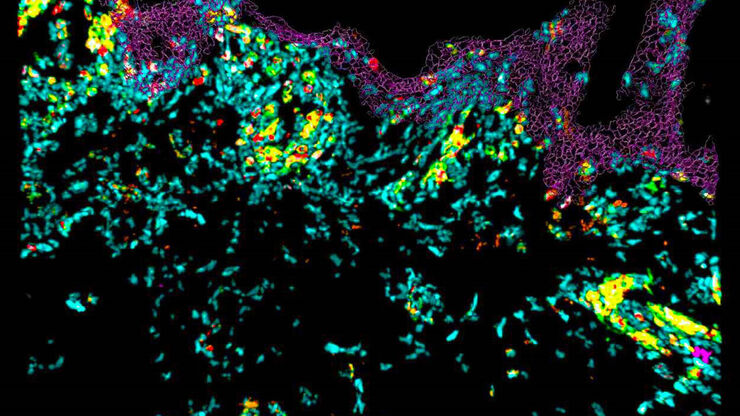
 and astrocytes (green) in a cortical spheroid derived from human induced pluripotent stem cells.")
活细胞成像指南
在生命科学各研究领域的广泛应用中,活细胞成像是一种不可或缺的工具,用于观察细胞在尽可能接近活体(即活的、活跃的)状态下的情况。本指南回顾了确保成功进行活细胞成像的各种重要注意事项,并介绍了各种旨在克服常见挑战的高性能解决方案。这些进展使我们能够对细胞生理学和动力学有新的认识。
Loading...
.")
利用光片显微技术聚焦三维长时程成像
长时程三维成像揭示了复杂的多细胞系统是如何生长和发育的,以及细胞是如何随着时间的推移而移动和相互作用的,从而揭示了发育、疾病和再生方面的重要知识。光片显微镜一次只照射样品的一个薄片,大大减少了光损伤,保护了样品的活性。这种温和的高速技术可在数小时甚至数天内提供清晰的体数据,使研究人员能够实时捕捉生物学的发展过程。
 and tubulin (magenta), acquired using Viventis Deep. Courtesy of Akanksha Jain, Treutlein Lab ETH-DBSSE Basel (Switzerland).")
Loading...

来捕捉发育动态的3D成像
本应用说明展示了研究人员如何成功利用 Viventis Deep 双视角光片显微镜探索3D多细胞模型(包括有机体、球形体和胚胎)的高分辨率长期成像,从而为发育生物学和疾病研究带来新的可能性。
Loading...
。红色标记区域显示未分化细胞区(尾芽),灰色阴影表示该组织生成的对应区域。上图:七鳃鳗;中图:猫鲨;下")
如何研究胚胎发育过程中的基因调控网络
请与 Andrea Boni 博士一起参加本次点播网络研讨会,探索光片显微镜如何彻底改变发育生物学。这种先进的成像技术可对三维样本进行高速、容积式实时成像,且光毒性极低。通过用户实例了解光片显微镜如何增强我们对肠道和大脑类器官发育的理解,并深入了解徕卡显微系统公司的 Viventis Deep 显微镜背后的技术及其在长期成像中的应用。
Loading...
.")
双视野光片显微镜,适用于大型多细胞系统
展示复杂多细胞系统的动态是生物学中的一个基本目标。为了应对在大型时空尺度上进行活体成像的挑战,作者在《自然·方法》杂志上发表的一篇论文中介绍了一种开放式多样本双视野光片显微镜。研究发现,Viventis LS2 Live显微镜在以单细胞分辨率成像大型样本方面取得了显著进展。