
Filter articles
标签
产品
Loading...
Predictive Service Prevents Downtime in Ghent
At the VIB BioImaging Core in Ghent, Belgium, researchers depend on Leica’s Stellaris 8 confocal microscope to explore the frontiers of biomedical science. When Leica’s RemoteCare system detected a…
Loading...
, Tropomyosin (cardiomyocytes, red) and GFP (primordial cardiac layer, green).")
显微镜中的荧光
荧光显微技术是一种特殊的光学显微镜技术。它利用的是荧光色素在一定波长的光激发下发光的能力。通过抗体染色或荧光蛋白标记,可以用这种荧光色素标记感兴趣的蛋白质。这样就可以确定单分子物种的分布、数量及其在细胞内的定位。此外,还可以进行共定位和相互作用研究,使用可逆结合染料(如 Ca2+ 和 fura-2)观察离子浓度,以及观察细胞的内吞和外吞过程。如今,利用荧光显微镜甚至可以对亚分辨率颗粒进行成像。
Loading...
 and astrocytes (green) in a cortical spheroid derived from human induced pluripotent stem cells.")
活细胞成像指南
在生命科学各研究领域的广泛应用中,活细胞成像是一种不可或缺的工具,用于观察细胞在尽可能接近活体(即活的、活跃的)状态下的情况。本指南回顾了确保成功进行活细胞成像的各种重要注意事项,并介绍了各种旨在克服常见挑战的高性能解决方案。这些进展使我们能够对细胞生理学和动力学有新的认识。
Loading...
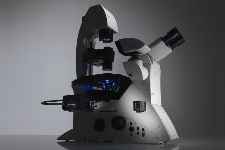
Factors to Consider When Selecting a Research Microscope
An optical microscope is often one of the central devices in a life-science research lab. It can be used for various applications which shed light on many scientific questions. Thereby the…
Loading...

线虫研究指南 - 针对线虫的相关工作
本指南概述了可以高效进行线虫的研究显微镜技术。线虫是一种广泛使用的模式生物,与人类有大约 70% 的基因同源性,是研究发育、神经科学、遗传学和衰老的理想生物。它的透明性和易培育性使其成为一个出色的遗传学模型系统。它可以进行高分辨率成像。主要的实验方法包括挑虫、转基因、荧光筛选、成像和记录。
Loading...

基于激光的视神经再生研究新方法
由于哺乳动物中枢神经系统(CNS)的自我修复能力有限以及传统损伤模型的不一致性,视神经再生是神经生物学的一大挑战。相比之下,爪蟾蝌蚪的视神经在受伤后可以再生,因此是研究轴突再生的分子和细胞机制的理想模型。在本应用说明中,我们展示了如何利用激光显微切割技术(LMD)对蝌蚪的视神经进行精确、一致的横切,从而开发出适合成像、转录组分析和功能恢复研究的高重复性损伤模型。
Loading...
 observed with an Ivesta 3 stereo microscope during fly pushing (sorting of the flies). The scale bar length is 1 mm. Image courtesy of M. Benton, EMBL, Heidelberg, Germany.")
Drosophila(果蝇)研究显微镜使用指南
一个多世纪以来,果蝇(典型的黑腹果蝇)一直被用作模式生物。原因之一是果蝇与人类共享许多与疾病相关的基因。果蝇经常被用于发育生物学、遗传学和神经科学的研究。果蝇的优点包括易于饲养且成本低廉、繁殖速度快、基因组完全测序以及可获得各种基因品系。使用徕卡显微镜可以进行高效的果蝇研究。

Loading...

前沿成像技术用于 GPCR 信号传导
通过这个按需网络研讨会,提升您的药理研究,了解 GPCR 信号传导,并探索旨在理解 GPCR 信号如何转化为细胞和生理反应的尖端成像技术。发现领先的研究,扩展我们对这些关键通路的认识,以寻找新的药物发现途径。
