显微镜知识库

显微镜知识库

显微镜知识库

徕卡显微系统的知识库提供有关显微镜学科的科学研究和教学材料。内容旨在对显微镜初学者、有经验的显微镜操作实践者和使用显微镜的科学家在他们的日常工作和实验有所帮助。这里有探索交互式教程和应用笔记,你可以找到你需要的显微镜的基础知识以及前沿技术——快来加入徕卡显微知识社区,分享您的专业知识!

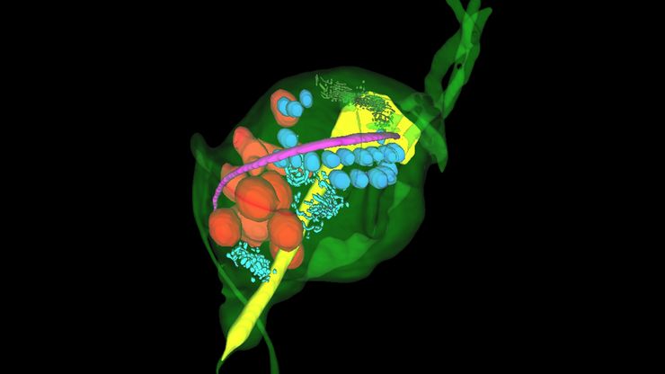
Integrated Serial Sectioning and Cryo-EM Workflows for 3D Biological Imaging

This on-demand webinar explores how integrated tools can support electron microscopy workflows from sample preparation to image analysis. Experts Andreia Pinto, Adrian Boey, and Hoyin Lai present the…
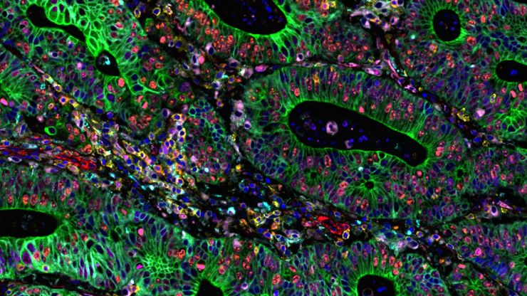
Cell DIVE multiplexed image of FFPE tissue section from human colon adenocarcinoma tissue.

Multiplexed Imaging Reveals Tumor Immune Landscape in Colon Cancer

Cancer immunotherapy benefits few due to resistance and relapse, and combinatorial therapeutic strategies that target multiple steps of the cancer-immunity cycle may improve outcomes. This study shows…
Cell DIVE multiplexed image of FFPE tissue section from human invasive ductal carcinoma (IDC)

AI-Powered Hi-Plex Spatial Analysis Tools for Breast Cancer Research

Breast cancer (BC) is the leading cause of cancer-related deaths in women. Investigating the tumor microenvironment (TME) is crucial to elucidate the mechanisms of tumor progression. Systematic…
Mouse brain slice which was immunostained with GFAP-A647 and imaged using a THUNDER Imager Tissue. Courtesy of H. Xu, University of Pennsylvania, Philadelphia, USA.

神经科学研究解决方案

您的工作是更好地了解神经退行性疾病,还是研究神经系统的功能? 了解如何使用徕卡显微系统的成像解决方案取得突破。
3D high-plex imaging in cancer immunology. Overview of a pancreatic tumor section in mouse model, labeled with 15 markers and imaged in one go using STELLARIS with SpectraPlex. (https://www.nature.com/articles/d42473-024-00260-7)

How to Streamline High-Plex Imaging for 3D Spatial Omics Advances

In this webinar, Dr. Julia Roberti and Dr. Luis Alvarez from Leica Microsystems introduce SpectraPlex, a new functionality integrated into the STELLARIS confocal platform for high-plex 3D spatial…
Pancreatic Ductal Adenocarcinoma with 11 Apoptosis biomarkers shown – BAK, BAX, BCL2, BCLXL, Caspase9, CIAP1, NaKATPase, PCK26, SMAC, Vimentin, and XIAP.

Transforming Research with Spatial Proteomics Workflows

Spatial Proteomics, Nature Methods 2024 Method of the Year, is driving research advancements in cancer, immunology, and beyond. By combining positional data with high throughput imaging of proteins in…
Large volume computational clearing processed Thunder image of human pancreatic islet organoid. Cells segmented using Segment By Example tool, automatically phenotyped, and color-coded based on phenotypes in Aivia. Image courtesy of the Matthias von Herrath Lab, La Jolla Institute of Immunology, La Jolla, CA.

利用人工智能图像分析工具更快、更轻松地获得洞察力

了解 Aivia 如何通过快速设置、准确的人工智能检测和简便的批量处理功能,帮助科学家简化图像分析。
Image: Human stem cell-derived mid brain organoids. Courtesy of Dr Tanya Singh, University of Oxford.

揭开类器官模型在生物医学研究中的秘密

准备深入了解类器官和3D培养物的世界,它们是促进我们了解人类健康的重要工具。浏览这些复杂的结构并获取清晰的图像进行分析是一项挑战。在本次活动中,来自牛津大学和伦敦大学学院的研究人员将与我们一起展示Thunder Imager Cell转盘共聚焦系统 如何提供更有说服力的高质量数据,以便深入了解各种模型。
Transfection using the Uncommon Bio reprogramming system. Image acquired using the THUNDER Imager 3D Cell Culture with THUNDER Large Volume Computational Clearing (LVCC) applied. Image courtesy of Samuel East, Uncommon Bio.

利用新型可扩展的干细胞培养设计未来

具有远见卓识的生物技术初创企业 Uncommon Bio 正在应对世界上最大的健康挑战之一:食品可持续性。在这次网络研讨会上,干细胞科学家塞缪尔-伊斯特(Samuel East)将展示他们如何使细胞农业的干细胞培养基既安全又经济可行。了解他们如何将培养基成本降低 1000 倍,并开发出不含动物成分、食品安全的 iPSC 培养基。
Scroll to top