Filter articles
标签
产品
Loading...

临床和手术显微镜在耳鼻喉科的应用
耳鼻喉科手术的数量逐年增加。 据估计,到2022年,全球每年进行的耳鼻喉科手术将超过2100万例。
Duane Mol医生是南非约翰内斯堡耳眼诊所的主任,同时也是该诊所的耳科医生和耳鼻喉科外科医生。此外,他还是国际耳鼻喉科网络LION的南非代表。
Loading...
 which is published by the US FDA (Food & Drug Administration).")
美国联邦法规第21章第11款和其他相关法规简介
本文概述了在美国(联邦法规第21章第11款)、欧盟(GMP附录11)和中国(NMPA)所用电子记录(数据输入、存储、签名和审批)的法规和指南,这些法规和指南会对医疗器械质量控制的数字化增强检测解决方案产生影响。与纸质记录方法相比,使用显微镜进行数字化增强检测具有更一致和更高效的检测优势。但是,与纸质记录和签名的规定相比,电子记录和签名的规定有明显不同的建议和要求。电子记录的创建、验证、存储和备份应…
Loading...

生理学图片库
生理学是关于生物体内的过程和功能。生理学研究的重点是生物体器官、组织或细胞的活动和功能,包括所涉及的物理和化学现象。在此,我们以不同的样本为例,向您展示与生理学有关的图片。
Loading...

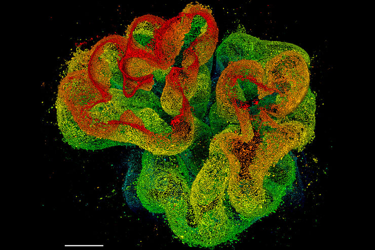
活细胞成像图库
活细胞显微镜技术是更好地了解细胞和分子功能的基础。如今,宽场显微镜是用于长时间观察细胞动态和发育的最常用技术。共聚焦显微镜也是一种重要工具,可生成三维结构图像,并以高空间和时间分辨率研究高度动态的细胞过程,同时使标本保持接近原生状态。
Loading...
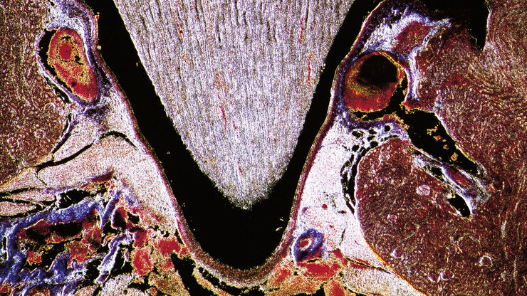
组织图片库
对动物和人体组织进行视觉分析对于了解癌症或神经变性等复杂疾病至关重要。从基本的免疫组化到体内成像,共聚焦显微镜和先进的模式可以让人们了解细胞、生物分子及其在环境中的相互作用。
 and astrocytes (green) in a cortical spheroid derived from human induced pluripotent stem cells.")
Loading...

多彩图库
荧光多色显微技术是多重成像技术的一个方面,可在同一实验中观察和分析同一样本中的多种元素--每种元素都标记有不同的荧光染料。这不仅能提高实验效率,还能获得更可靠、更有意义的结果,从而了解细胞和组织内的复杂过程。本图集展示了使用THUNDER和STELLARIS平台获得的标有多种荧光探针的样本图像。
Loading...
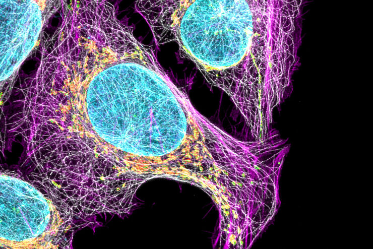
细胞生物学图片库
细胞生物学研究细胞的结构、功能和行为,包括细胞新陈代谢、细胞周期和细胞信号传导。荧光显微镜是细胞生物学家工具包中不可或缺的一部分。宽场显微镜和共聚焦显微镜被广泛用于观察细胞内复杂结构的细节。