显微镜知识库

显微镜知识库

显微镜知识库

徕卡显微系统的知识库提供有关显微镜学科的科学研究和教学材料。内容旨在对显微镜初学者、有经验的显微镜操作实践者和使用显微镜的科学家在他们的日常工作和实验有所帮助。这里有探索交互式教程和应用笔记,你可以找到你需要的显微镜的基础知识以及前沿技术——快来加入徕卡显微知识社区,分享您的专业知识!
C. elegans embedded in Lowicryl® HM20; pharynx showing red fluorescence (mCherry). The overview shows a front view onto the resin capsule formed by the bottom of a flow-through chamber of the EM AFS2. The capsule was pretrimmed manually. The blockface was trimmed automatically using the AutoTrim function of UC Enuity guided by fluorescence of the worm. Edge length of both squares in relation to the images is 250 µm.

How Fluorescence Guides Sectioning of Resin-embedded EM Samples

Electron microscopes, including transmission electron microscopes (TEM) and scanning electron microscopes (SEM), are widely utilized to gain detailed structural information about biological samples or…

Coherent Raman Scattering Microscopy Publication List

CRS (Coherent Raman Scattering) microscopy is an umbrella term for label-free methods that image biological structures by exploiting the characteristic, intrinsic vibrational contrast of their…
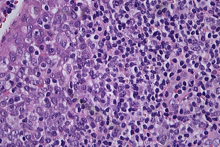
Histopathological sample, 40x magnification

临床显微镜:相机选择的考虑因素

过去几年,病理学实验室对图像的需求显著增加,无论是在组织病理学、细胞学、血液学、临床微生物学还是其他应用领域。除了诊断记录外,图像还服务于许多其他目的。然而,通过目镜观察到的图像和数字图像在本质上是不同的,一个是光学图像,另一个是数字图像。从与相机相关的几个方面来审视这一过程,将有助于确保您能够获取所有细节和颜色保真度的图像。
Visoria B

选择临床显微镜需考虑的因素

显微镜是病理学家工作流程中不可或缺的一部分。特别是在组织病理学、血液病理学或医学微生物学中,病理学家使用显微镜来高效、可靠地进行诊断。因此,他们经常长时间低头看目镜,可能会因为工作姿势而感到身体不适。
Multiplexed Cell DIVE imaging of Adult Human Alzheimer’s Brain Tissue labelled with 15 antibodies targeted towards markers specific to astrocytes (GFAP, S100B), microglia (TMEM119, IBA1), and Alzheimer’s-associated markers (β-amyloid and p-Tau217).

利用大数据探索阿尔茨海默病的空间蛋白组

阿尔茨海默病是一种遗传性和散发性的神经退行性疾病,导致中晚年认知能力下降,特征为β-淀粉样蛋白斑块和 tau蛋白 缠结。由于治疗选择有限,新的研究策略至关重要。Cell DIVE 多重成像解决方案可以对阿尔茨海默病脑组织进行研究,揭示,可能新的研究方向。这里我们展示了 Cell DIVE 多重成像仪的图像查看器,用户能够直接在自己的浏览器中访问完整的阿尔茨海默病多重数据集。
Colon adenocarcinoma and normal colon at the tumor margin. 13 biomarkers shown including Cadherin, CD3, CD4, CD8, CD20, CD31, CD45, Collagen, Caspase 9, BCL2, Beta-Catenin, Vimentin, and Smooth Muscle Actin.

利用大数据查看器揭示结肠癌隐藏的复杂性

结直肠癌是一种的重大健康负担。虽然手术初期有效,但部分患者会发展为预后不良的复发性继发疾病,需要采用免疫疗法等先进治疗手段。利用空间生物学方法,如 Cell DIVE 多重成像技术,可为开发新型治疗方案提供关键洞见。通过 Minerva 图像查看器在浏览器中访问完整的 Cell DIVE 数据集,进一步探索这些发现。
Pancreatic Ductal Adenocarcinoma with 11 Aerobic Glycolysis/Warburg Effect biomarkers shown – BCAT, Glut1, HK2, HTR2B, LDHA, NaKATPase, PCAD, PCK26, PKM2, SMA1, and Vimentin.

用大数据视角深入了解胰腺癌研究

胰腺癌由于其靠近主要器官难以分辨和难治疗,死亡率接近 40%,。这个研究探讨了胰腺导管腺癌(PDAC)的复杂生物学机制,研究了代谢、凋亡和免疫中肿瘤侵袭性的相关分子结构和空间决定因素。可以访问您的浏览器中的完整 Cell DIVE 数据集,以深入了解这些发现。
Blood vessel system of a zebrafish larvae

克服显微镜成像移动斑马鱼幼虫时的挑战

Zebrafish is a valuable model organism with many beneficial traits. However, imaging a full organism poses challenges as it is not stationary. Here, this case study shows how zebrafish larvae can be…
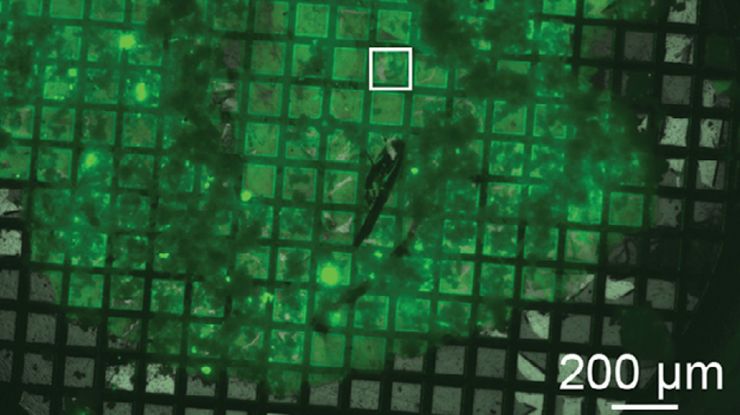
Block-face created by automatic trimming under fluorescence. Mammalian cells of interest, stained with CellTrackerTM Green are visualized within the block-face using the UC Enuity equipped with the stereo microscope M205 FA. In the background a carbon finder grid in black is visible. All samples in the article are created by Felix Gaedke, PhD, CECAD, Cologne, Germany.

如何在块面中自动获取感兴趣的荧光细胞

本文介绍了使用超薄切片超薄切片机自动修整修块功能,获取树脂块面中带有荧光信号的细胞结构。我们展示了如何使用配置有体视显微镜 M205 FA 的超薄切片超薄切片机 UC Enuity ,来识别感兴趣的荧光细胞,如何自动修整包含细胞的块面,以及如何在切片中观察细胞而无需转移到外部显微镜。
Scroll to top