
Filter articles
标签
产品
Loading...
Computational Clearing - 增强3D标本成像
本次网络研讨会旨在阐明有助于THUNDER显微成像仪实现三维样品可视化的关键规格,并改进研究人员的成像相关工作流程。
Loading...
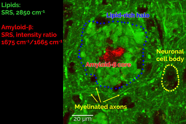
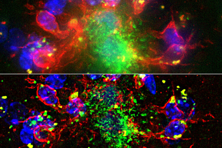
受激拉曼散射显微镜探测神经退行性疾病
Despite decades of research, the molecular mechanisms underlying some of the most severe neurodegenerative diseases, such as Alzheimer’s or Parkinson’s, remain poorly understood. The progression of…
Loading...

斑马鱼大脑高分辨率全器官成像
结构信息是理解复杂生物系统的关键,而脊椎动物的中枢神经系统是最复杂的生物结构之一。要想从发育中的斑马鱼身上分离出一个完整的大脑,我们需要覆盖大约10平方毫米的区域,深度在毫米范围内。通常,低倍透镜不能提供足够的分辨率来揭示神经组织中复杂结构之间的相互作用。此外,由于散射过程,使用共聚焦显微镜在致密生物组织内成像深度通常限制在大约10微米。
Loading...

使用旋转设备进行光片样本安装
TCS SP8 DLS 显微镜采用了一种创新的设计理念,将光片显微技术集成到共聚焦显微镜中。得益于其独特的光学架构,样本可以像进行标准共聚焦成像一样,安装在标准玻璃底培养皿上。与传统的样本安装程序相比,这一过程只需进行少量调整。
Loading...
DIVE 多光子显微镜图像库
当今的生命科学研究集中于复杂的生物过程,例如癌症和其他人类疾病的原因。深入观察组织和活体标本对于理解细胞中的条件和机制以及寻找生命科学中面临的关键问题的答案至关重要。

Loading...

使用 U 形玻璃毛细管进行样品装载
徕卡显微系统的DLS显微镜系统是一种创新概念,将光片显微技术集成到共聚焦平台中。由于其独特的光学结构,样本可以安装在标准玻璃底培养皿上,与传统的安装程序相比,几乎不需要或只需很少的适应。在这里,我们介绍了一种便捷的方法,能够快速准备样本以进行光片成像。
Loading...

不可能的任务:可调颜色用于非扫描检测
徕卡显微系统的 4Tune 探测器是 SP8 DIVE 深度体内探测器的关键组件,提供具有非扫描检测的光谱可调图像记录,是多参数多光子显微镜的创新解决方案。
Loading...
 stereo microscope for a task like surgery.")
Rodent and Small-Animal Surgery
Learn how you can perform rodent (mouse, rat, hamster) and small-animal surgery efficiently with a microscope for developmental biology and medical research applications by reading this article.
