
Filter articles
标签
产品
Loading...
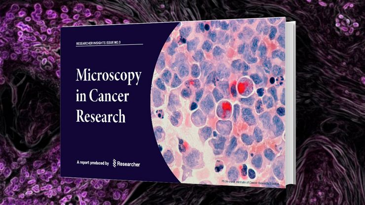
Researchers Insights: Microscopy in Cancer Research
Discover how imaging techniques are driving cancer research forward. In this issue, we present comprehensive multimodal studies using microscopy, as well as new directions in intraoperative cancer…
Loading...
, Tropomyosin (cardiomyocytes, red) and GFP (primordial cardiac layer, green).")
显微镜中的荧光
荧光显微技术是一种特殊的光学显微镜技术。它利用的是荧光色素在一定波长的光激发下发光的能力。通过抗体染色或荧光蛋白标记,可以用这种荧光色素标记感兴趣的蛋白质。这样就可以确定单分子物种的分布、数量及其在细胞内的定位。此外,还可以进行共定位和相互作用研究,使用可逆结合染料(如 Ca2+ 和 fura-2)观察离子浓度,以及观察细胞的内吞和外吞过程。如今,利用荧光显微镜甚至可以对亚分辨率颗粒进行成像。
Loading...
 and astrocytes (green) in a cortical spheroid derived from human induced pluripotent stem cells.")
活细胞成像指南
在生命科学各研究领域的广泛应用中,活细胞成像是一种不可或缺的工具,用于观察细胞在尽可能接近活体(即活的、活跃的)状态下的情况。本指南回顾了确保成功进行活细胞成像的各种重要注意事项,并介绍了各种旨在克服常见挑战的高性能解决方案。这些进展使我们能够对细胞生理学和动力学有新的认识。
Loading...
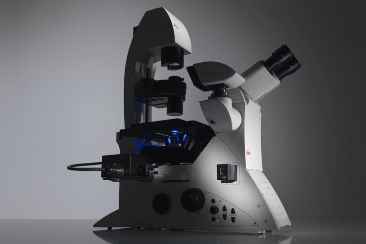
Factors to Consider When Selecting a Research Microscope
An optical microscope is often one of the central devices in a life-science research lab. It can be used for various applications which shed light on many scientific questions. Thereby the…
 and tubulin (magenta), acquired using Viventis Deep. Courtesy of Akanksha Jain, Treutlein Lab ETH-DBSSE Basel (Switzerland).")
Loading...
 at 2 weeks. Image acquired using Mica.")
如何为深层肌肉组织中的轴突再生成像
这项研究重点介绍了亚伦-李(Aaron Lee)博士对截肢后肌肉移植中神经再生的定位研究。肢体缺失通常会导致生活质量下降,这不仅是因为组织缺失,还因为轴突再生紊乱引起的神经性疼痛。Mica组织学成像和荧光成像可帮助了解神经再生过程中轴突的生长和分支这项研究有助于塑造未来的神经假体接口设计,改善患者的治疗效果。
Loading...
 observed with an Ivesta 3 stereo microscope during fly pushing (sorting of the flies). The scale bar length is 1 mm. Image courtesy of M. Benton, EMBL, Heidelberg, Germany.")
Drosophila(果蝇)研究显微镜使用指南
一个多世纪以来,果蝇(典型的黑腹果蝇)一直被用作模式生物。原因之一是果蝇与人类共享许多与疾病相关的基因。果蝇经常被用于发育生物学、遗传学和神经科学的研究。果蝇的优点包括易于饲养且成本低廉、繁殖速度快、基因组完全测序以及可获得各种基因品系。使用徕卡显微镜可以进行高效的果蝇研究。

Loading...

Mica: 助力伦敦帝国学院开展跨学科科研研究
这篇访谈重点介绍了伦敦帝国学院的 Mica 所产生的变革性影响。科学家们解释了Mica如何改变了游戏规则,扩大了研究的可能性,促进了跨学科合作。他们解释了使用 Mica 进行详细的活细胞成像如何提供更有意义的信息,使科学家始终站在研究的最前沿。研究小组预计,Mica将继续开辟新的研究途径,包括研究微流体技术和其他先进应用。
