Filter articles
标签
产品
Loading...
.")
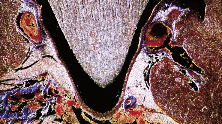
利用激光显微切割(LMD)在空间背景下分离神经元
在阿尔茨海默病之后,帕金森病是第二常见的进行性神经退行性疾病。在首发症状出现之前,中脑中高达70%的多巴胺释放神经元已经死亡。本文描述了如何使用现代激光显微切割(LMD)方法帮助解决帕金森病之谜。研究涉及在空间背景下分离和分析神经元。这些细胞来自帕金森病患者的死后黑质组织样本,以便深入了解该病的分子机制。
Loading...

激光显微切割技术如何助力神经科学研究取得开创性进展?
玛尔塔·帕特林尼博士,卡罗林斯卡学院的高级科学家,分享了她在成人人类神经发生开创性研究中使用激光显微切割(LMD)的经验,并提供了关于LMD在空间蛋白质组学和精准医学中未来应用潜力的个人见解。
Loading...

激光显微切割技术用于组织和细胞分离的协议 - 免费下载电子书
激光显微切割(LMD,也称为激光捕获显微切割或LCM)使用户能够分离特定的单个细胞或整个组织区域,甚至亚细胞结构如染色体。纯化的组织和细胞可用于下游的RNA、DNA和蛋白质组工作流程。

Loading...

高效清洁度分析的关键因素
在汽车和电子行业,零部件上细小的污染颗粒物也可能影响产品的性能,导致产品出现故障,或使用寿命缩短。对于汽车来说,过滤系统很容易受到影响。对于电子产品来说,印刷电路板(PCB)或连接器上的污染可能会导致短路。因此,清洁度在现代制造业的质量控制中占有核心地位,特别是使用由不同供应商生产的部件时,更要重点关注清洁情况。车辆或设备的关键部件如果受到污染,整个系统就可能发生故障。因此,高效清洁度分析过程必须…