显微镜知识库

显微镜知识库

显微镜知识库

徕卡显微系统的知识库提供有关显微镜学科的科学研究和教学材料。内容旨在对显微镜初学者、有经验的显微镜操作实践者和使用显微镜的科学家在他们的日常工作和实验有所帮助。这里有探索交互式教程和应用笔记,你可以找到你需要的显微镜的基础知识以及前沿技术——快来加入徕卡显微知识社区,分享您的专业知识!
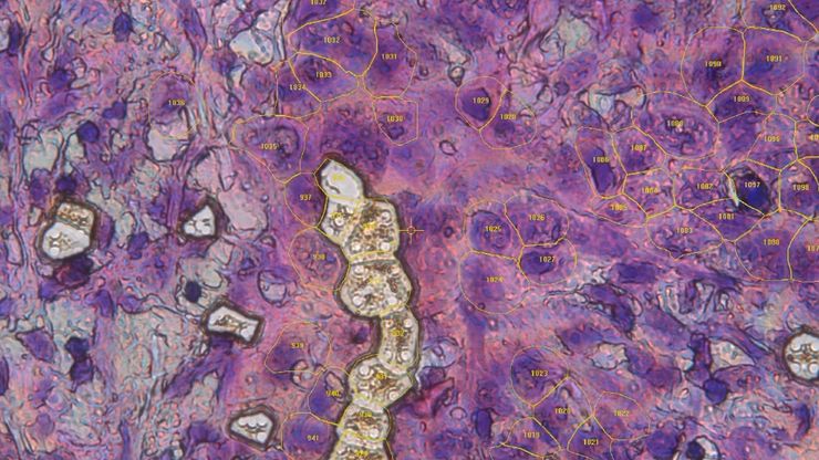
Single cells collected via laser microdissection as part of the Deep Visual Proteomics workflow.

AI meets Deep Visual Proteomics (DVP) to Advance Disease Research

In this webinar, Dr. Andreas Mund introduces Deep Visual Proteomics (DVP) – a cutting-edge platform that integrates AI-powered tissue modeling with spatially resolved, untargeted proteomics. He…
这些图像说明,要捕捉特定细胞中的所有 gH2Ax 病灶并进行精确计数,用多个三维光切片方法实现。

罕见疾病 CRISPR 疗法的开发与风险解除

Fyodor Urnov博士和Sadik Kassim博士最初是在ASGCT 2025会议上作这一按需演讲的,演讲的重点是遗传医学中的一个关键挑战:如何将CRISPR疗法从单一疾病解决方案扩展到平台方法,特别是针对罕见的儿科遗传疾病。Urnov 博士展示了由 Matthew Kan 博士领导的创新基因组研究所的工作,这是 IGI-Danaher Beacon for CRISPR Cures…

用于三维生物成像的集成连续切片与冷冻电镜工作流程

本场网络研讨会探讨了集成化工具如何支持从样品制备到图像分析的电子显微镜全流程。专家Andreia Pinto博士、Adrian Boey博士与Hoyin Lai博士将介绍UC Enuity超薄切片机和Aivia图像分析平台,并演示这些工具如何同时适用于常温与低温实验环境。会议内容包含阵列断层成像、基于深度学习的图像分割、以及生物成像中cryo-lift-out工作流程的实际案例解析。
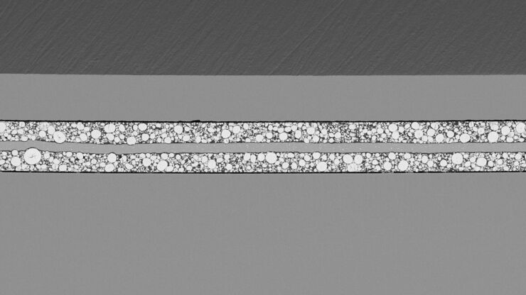
SEM image of the full Li-NMC electrode sample, showing the two porous layers and the metal film at the center of the structure.

通过Cryo-EM(冷冻电镜)和 CryoFIB(冷冻聚焦离子束) 揭示钠电池退化机制

探索低温电镜和聚焦离子束技术如何揭示钠电池界面的内在结构。本次研讨会将提出基于隔膜渗透(而非枝晶生长)的新型退化模型,并解析电解液溶剂如何影响界面稳定性与电池性能。
3D high-plex imaging in cancer immunology. Overview of a pancreatic tumor section in mouse model, labeled with 15 markers and imaged in one go using STELLARIS with SpectraPlex. (https://www.nature.com/articles/d42473-024-00260-7)

如何优化多标成像技术推动3D空间组学发展

本次网络研讨会上,徕卡显微系统的Julia Roberti博士与Luis Alvarez博士将介绍STELLARIS共聚焦平台的全新功能SpectraPlex,该技术可实现超多标三维空间成像。该技术旨在通过实现超多标成像且无需频繁人工干预,从而简化和增强空间生物学应用。
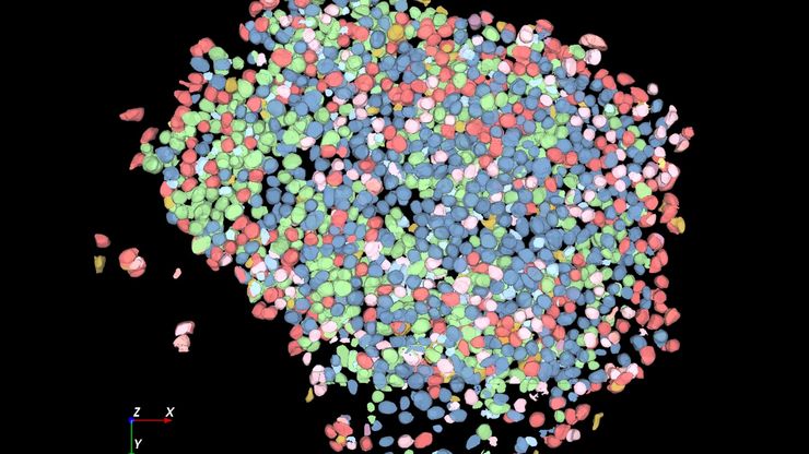
Large volume computational clearing processed Thunder image of human pancreatic islet organoid. Cells segmented using Segment By Example tool, automatically phenotyped, and color-coded based on phenotypes in Aivia. Image courtesy of the Matthias von Herrath Lab, La Jolla Institute of Immunology, La Jolla, CA.

利用人工智能图像分析工具更快、更轻松地获得洞察力

了解 Aivia 如何通过快速设置、准确的人工智能检测和简便的批量处理功能,帮助科学家简化图像分析。
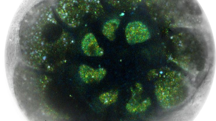
Image: Human stem cell-derived mid brain organoids. Courtesy of Dr Tanya Singh, University of Oxford.

揭开类器官模型在生物医学研究中的秘密

准备深入了解类器官和3D培养物的世界,它们是促进我们了解人类健康的重要工具。浏览这些复杂的结构并获取清晰的图像进行分析是一项挑战。在本次活动中,来自牛津大学和伦敦大学学院的研究人员将与我们一起展示Thunder Imager Cell转盘共聚焦系统 如何提供更有说服力的高质量数据,以便深入了解各种模型。
Transfection using the Uncommon Bio reprogramming system. Image acquired using the THUNDER Imager 3D Cell Culture with THUNDER Large Volume Computational Clearing (LVCC) applied. Image courtesy of Samuel East, Uncommon Bio.

利用新型可扩展的干细胞培养设计未来

具有远见卓识的生物技术初创企业 Uncommon Bio 正在应对世界上最大的健康挑战之一:食品可持续性。在这次网络研讨会上,干细胞科学家塞缪尔-伊斯特(Samuel East)将展示他们如何使细胞农业的干细胞培养基既安全又经济可行。了解他们如何将培养基成本降低 1000 倍,并开发出不含动物成分、食品安全的 iPSC 培养基。
Complete camera overview of EM grid recorded with 3 channels. Inserts displaying the positions, where superresolved 3D confocal images were recorded. 3D renderings of these positions are shown in the zoomed inserts. Fluorescence channels (nuclei by Hoechst, blue; mitochondria by MitoTracker Green, green; lipid Droplets by Bodipy and Crimson Beads, red). Width of a grid square is 90 ?m, width of a grid bar is 35 ?m. Samples kindly provided by Ievgeniia Zagoriy, Mahamid-Group, EMBL Heidelberg, Germany.

从显微镜到电镜:完整的冷冻光电联用工作流程

在题为“多模态玻璃化征程,从实验台到电子显微镜的冷冻关联工作流程”的网络研讨会上,专家团队(Edoardo D'Imprima、Zhengyi Yang、Andreia Pinto 和 Martin…
Scroll to top