
Filter articles
标签
产品
Loading...
 and astrocytes (green) in a cortical spheroid derived from human induced pluripotent stem cells.")
Guide to Live-Cell Imaging
For a wide range of applications in various research fields of life science, live-cell imaging is an indispensable tool for visualizing cells in a state as close to in vivo, i.e. living and active, as…
Loading...

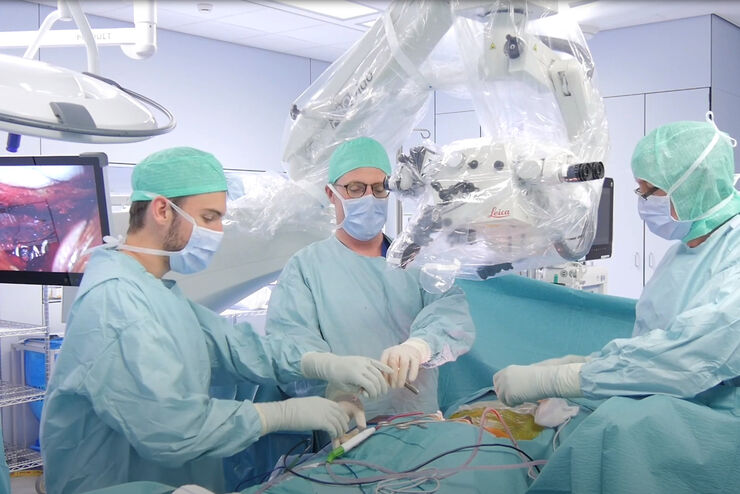
神经外科和眼科中的融合光学 - 更大三维聚焦区域
神经外科医生和眼科医生处理精细结构、深或狭窄的腔体以及具有至关重要功能的微小结构。因此,手术区域的清晰三维视图对手术结果和患者安全至关重要。到目前为止,增加景深以获得更大三维聚焦区域只能通过降低分辨率来实现。一项新技术能够克服这一挑战。
Loading...
 image of a cross section of C. elegans (roundworm). Courtesy of T. Müller-Reichert, MPI-CBG, Dresden, Germany and K. McDonald, University of California, Berkeley, USA.")
高压冷冻简介
水是细胞最主要的组成部分,因此对于维持细胞超微结构至关重要。目前,冷冻固定是固定细胞成分,而不导致其显著结构变化的唯一途径。现阶段有两种常见的方法:投入冷冻与高压冷冻固定。
Loading...
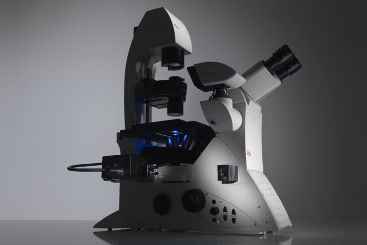
Factors to Consider When Selecting a Research Microscope
An optical microscope is often one of the central devices in a life-science research lab. It can be used for various applications which shed light on many scientific questions. Thereby the…
Loading...
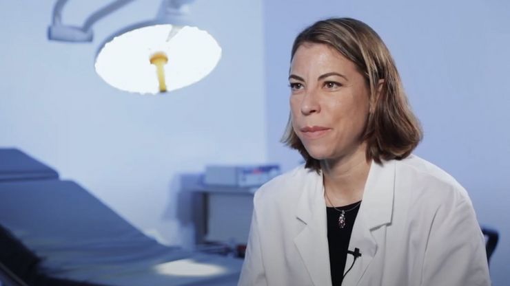
颌面整形与重建手术中的先进可视化技术
整形与重建手术要求极高。手术显微镜发挥着重要作用,能确保皮瓣血管化良好。
Dr. Christine Bach 是法国 Suresnes 地区 Foch 医院的整形与重建外科医生,专攻头颈部手术,为耳鼻喉癌症患者实施重建手术。

Loading...

无限远光学系统
“无限远光学”这一概念是指在显微镜的物镜和镜筒透镜之间具有平行光线的光束路径。平面光学元件可以进入到这个“无限远空间”中,而不影响成像,这对于利用DIC或荧光等对比度方法至关重要。
现代显微技术需要在无限远光路中添加多种光学仪器,如光源或激光装置。满足这一需求的不同方法已经出现,本文对其进行了描述。
Loading...

跨行业的质量保证改进
精确是最重要的。试想一下,心脏起搏器在运行过程中发生故障,或者半导体缺陷导致关键系统崩溃。在医疗设备、电子产品和半导体等行业,误差几乎为零。质量保证(QA)不再仅仅是一项监管要求,而是一项推动业务成功和保护品牌完整性的战略优势。
