
Filter articles
标签
产品
Loading...
 and astrocytes (green) in a cortical spheroid derived from human induced pluripotent stem cells.")
Guide to Live-Cell Imaging
For a wide range of applications in various research fields of life science, live-cell imaging is an indispensable tool for visualizing cells in a state as close to in vivo, i.e. living and active, as…
Loading...
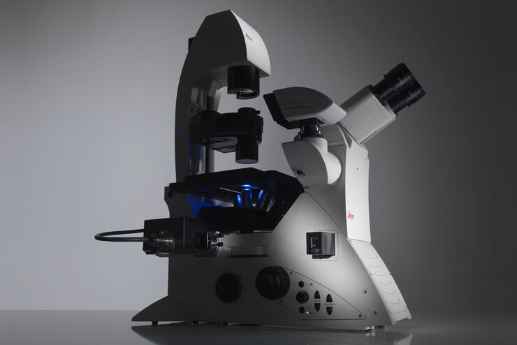
Factors to Consider When Selecting a Research Microscope
An optical microscope is often one of the central devices in a life-science research lab. It can be used for various applications which shed light on many scientific questions. Thereby the…
Loading...

无限远光学系统 - 从 "无限远光学 "到无限远端口
"无限远光学 "是指显微镜的物镜和管状透镜之间的光路具有平行光线的概念 [1]。在这个 "无限远空间 "中放置平面光学元件不会影响图像的形成,这对于科学应用中常用的 DIC 或荧光等对比方法至关重要。需要在无限远光路中添加光源或激光设备等仪器。本文介绍了满足这一需求的不同方法。
 and tubulin (magenta), acquired using Viventis Deep. Courtesy of Akanksha Jain, Treutlein Lab ETH-DBSSE Basel (Switzerland).")
Loading...
 at Leica Microsystems.")
空间蛋白质组学的突破如何拯救生命
中毒性表皮坏死溶解症(TEN)是一种罕见的、但对抗生素或痛风治疗等常见药物的破坏性反应。这种疾病开始时并无大碍,通常只是皮疹,但会迅速升级为大面积皮肤脱落,类似于严重烧伤。尽管 TEN病情十分严重,但其基本机制仍然难以捉摸,治疗方案也仅限于支持性护理。TEN 的死亡率高达 30%,长期以来一直是临床医生的噩梦,直到现在才有了靶向疗法。
Loading...

基于激光的视神经再生研究新方法
由于哺乳动物中枢神经系统(CNS)的自我修复能力有限以及传统损伤模型的不一致性,视神经再生是神经生物学的一大挑战。相比之下,爪蟾蝌蚪的视神经在受伤后可以再生,因此是研究轴突再生的分子和细胞机制的理想模型。在本应用说明中,我们展示了如何利用激光显微切割技术(LMD)对蝌蚪的视神经进行精确、一致的横切,从而开发出适合成像、转录组分析和功能恢复研究的高重复性损伤模型。
Loading...
 at 2 weeks. Image acquired using Mica.")
如何为深层肌肉组织中的轴突再生成像
这项研究重点介绍了亚伦-李(Aaron Lee)博士对截肢后肌肉移植中神经再生的定位研究。肢体缺失通常会导致生活质量下降,这不仅是因为组织缺失,还因为轴突再生紊乱引起的神经性疼痛。Mica组织学成像和荧光成像可帮助了解神经再生过程中轴突的生长和分支这项研究有助于塑造未来的神经假体接口设计,改善患者的治疗效果。

Loading...

利用快速高对比度成像改进斑马鱼-胚胎筛查
通过这篇文章,您可以了解如何利用 DM6 B 显微镜的高速、高对比度成像技术促进转基因斑马鱼胚胎的筛选,从而确保发育生物学研究的准确定位。
