
Filter articles
主题和标签
产品
Loading...
.")
AI-Powered Hi-Plex Spatial Analysis Tools for Breast Cancer Research
Breast cancer (BC) is the leading cause of cancer-related deaths in women. Investigating the tumor microenvironment (TME) is crucial to elucidate the mechanisms of tumor progression. Systematic…

Loading...

The “Waffle Method”: High-Pressure Freeze Complex Samples
This article describes the advantages of a special high pressure freezing method, the so-called “Waffle Method”. Learn how the “Waffle Method” uses EM grids as spacers for high-pressure freezing,…
Loading...
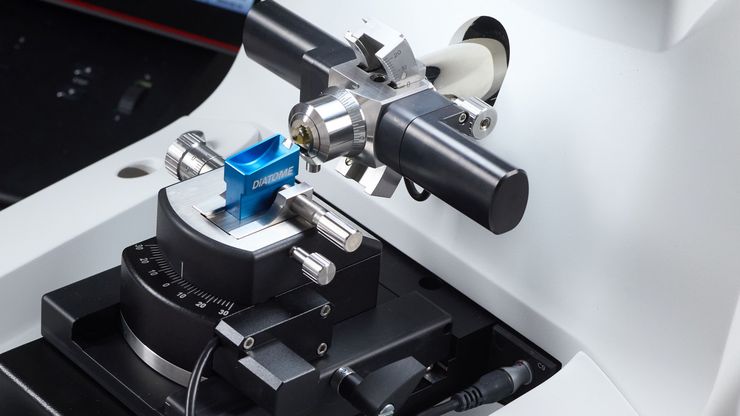
Mastering Polymer Sectioning with Helmut Gnaegi
When it comes to ultramicrotomy, few names carry the weight of Helmut Gnaegi. As co-founder of Diatome, a global leader in diamond knife technology, Helmut has spent decades refining the art and…
Loading...
斑马鱼研究
为了在筛选、分拣、操作和成像过程中获取高质量结果,您需要观察细节和结构,从而为您的下一步研究做出正确的决策。
徕卡体视显微镜和透射光底座以出众的光学器件和优良的分辨率而闻名,是全世界研究学者的首选。
Loading...
")
How to Streamline High-Plex Imaging for 3D Spatial Omics Advances
In this webinar, Dr. Julia Roberti and Dr. Luis Alvarez from Leica Microsystems introduce SpectraPlex, a new functionality integrated into the STELLARIS confocal platform for high-plex 3D spatial…
Loading...

The Guide to Augmented Reality in Microsurgery
In an era of technological advancement, Augmented Reality (AR) is rapidly transforming the medical field. In surgical microscopy, AR can display fluorescence signals as digital overlays in real-time…
Loading...

Improving Zebrafish-Embryo Screening with Fast, High-Contrast Imaging
Discover from this article how screening of transgenic zebrafish embryos is boosted with high-speed, high-contrast imaging using the DM6 B microscope, ensuring accurate targeting for developmental…
Loading...

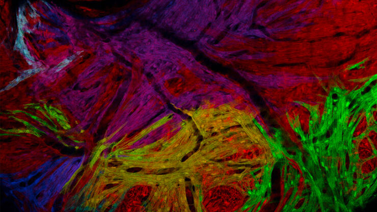
Transforming Research with Spatial Proteomics Workflows
Spatial Proteomics, Nature Methods 2024 Method of the Year, is driving research advancements in cancer, immunology, and beyond. By combining positional data with high throughput imaging of proteins in…
