
Filter articles
标签
产品
Loading...
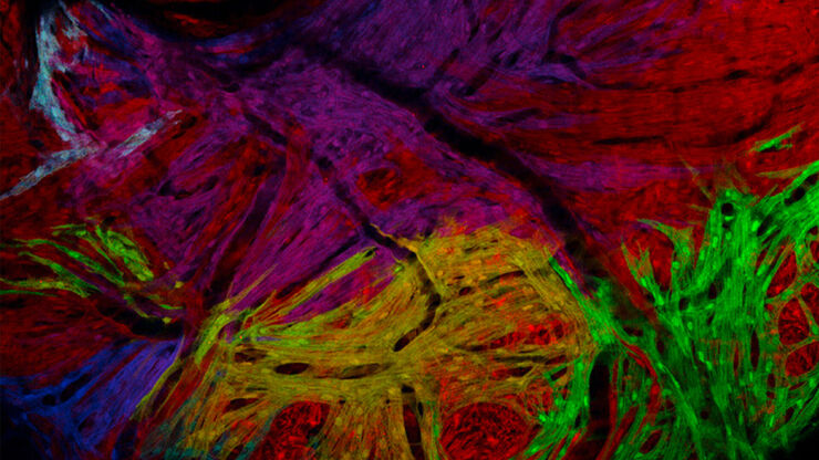
, Tropomyosin (cardiomyocytes, red) and GFP (primordial cardiac layer, green).")
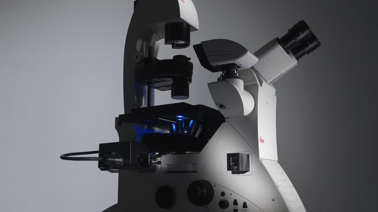
显微镜中的荧光
荧光显微技术是一种特殊的光学显微镜技术。它利用的是荧光色素在一定波长的光激发下发光的能力。通过抗体染色或荧光蛋白标记,可以用这种荧光色素标记感兴趣的蛋白质。这样就可以确定单分子物种的分布、数量及其在细胞内的定位。此外,还可以进行共定位和相互作用研究,使用可逆结合染料(如 Ca2+ 和 fura-2)观察离子浓度,以及观察细胞的内吞和外吞过程。如今,利用荧光显微镜甚至可以对亚分辨率颗粒进行成像。
Loading...
 and astrocytes (green) in a cortical spheroid derived from human induced pluripotent stem cells.")
活细胞成像指南
在生命科学各研究领域的广泛应用中,活细胞成像是一种不可或缺的工具,用于观察细胞在尽可能接近活体(即活的、活跃的)状态下的情况。本指南回顾了确保成功进行活细胞成像的各种重要注意事项,并介绍了各种旨在克服常见挑战的高性能解决方案。这些进展使我们能够对细胞生理学和动力学有新的认识。
Loading...
选择研究用显微镜时应考虑的因素
光学显微镜通常是生命科学研究实验室的核心设备之一。它可用于各种应用,揭示许多科学问题。因此,显微镜的配置和功能对其应用范围至关重要,从明视野显微镜到荧光显微镜,再到活细胞成像。本文简要概述了显微镜的相关功能,并总结了在选择研究用显微镜时应考虑的关键问题。
Loading...

线虫研究指南 - 针对线虫的相关工作
本指南概述了可以高效进行线虫的研究显微镜技术。线虫是一种广泛使用的模式生物,与人类有大约 70% 的基因同源性,是研究发育、神经科学、遗传学和衰老的理想生物。它的透明性和易培育性使其成为一个出色的遗传学模型系统。它可以进行高分辨率成像。主要的实验方法包括挑虫、转基因、荧光筛选、成像和记录。
Loading...
 observed with an Ivesta 3 stereo microscope during fly pushing (sorting of the flies). The scale bar length is 1 mm. Image courtesy of M. Benton, EMBL, Heidelberg, Germany.")
Drosophila(果蝇)研究显微镜使用指南
一个多世纪以来,果蝇(典型的黑腹果蝇)一直被用作模式生物。原因之一是果蝇与人类共享许多与疾病相关的基因。果蝇经常被用于发育生物学、遗传学和神经科学的研究。果蝇的优点包括易于饲养且成本低廉、繁殖速度快、基因组完全测序以及可获得各种基因品系。使用徕卡显微镜可以进行高效的果蝇研究。

Loading...
斑马鱼研究指南
在斑马鱼研究过程中,尤其是在筛选、分类、处理和成像过程中,要想获得最佳结果,看到精细的细节和结构非常重要。他们帮助研究人员为下一步做出正确的决定。徕卡体视显微镜以出色的光学性能和分辨率著称,配备透射光基底和荧光照明,为斑马鱼成像提供了合适的解决方案。高分辨率、色彩保真度和最佳对比度使研究人员能够做出具有洞察力的决策。
Loading...

显微外科增强现实技术指南
在技术进步的时代,增强现实技术(AR)正在迅速改变医疗领域。在手术显微镜中,AR 可以在手术视野内以数字叠加的方式实时显示荧光信号,为外科医生提供有关荧光区域的更多信息。外科医生的视图可通过不同系统的数据叠加来增强,提供叠加到实时显微镜视图上的功能或解剖信息。

