Filter articles
标签
Loading...
. With DIC users are able to visualize small height differences on the wafer surface more easily.")
6 英寸晶片检测显微镜,可靠的观察微小高度差
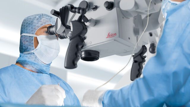
本文介绍了一种 6 英寸晶圆检测显微镜,无论用户的技术水平如何,它都能自动进行可重复的 DIC(微分干涉对比)成像。集成电路 (IC) 芯片和半导体元件的制造需要晶圆检测,以确保不存在影响性能的缺陷。通常使用光学显微镜进行质量控制、故障分析和 R&D 检测。为了有效地观察晶圆上结构之间的微小高度差,可以使用 DIC。
Loading...
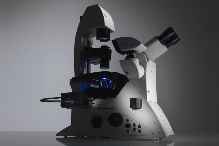
Factors to Consider When Selecting a Research Microscope
An optical microscope is often one of the central devices in a life-science research lab. It can be used for various applications which shed light on many scientific questions. Thereby the…
 and oblique (right) brightfield illumination using a Leica compound microscope. The defect on the wafer surface is clearly more visible with oblique illuminati")

Loading...

哺乳动物细胞培养的介绍
哺乳动物细胞培养是生命科学的基本支柱之一。如果不具备在实验室中培养细胞的能力,那么细胞生物学、免疫学、肿瘤研究等学科很难实现快速发展。本文概述了哺乳动物细胞培养系统,可以根据其形态、细胞类型和组织对其进行分类。此外,还介绍了适宜的细胞生长条件以及需要使用何种显微镜来观察细胞。
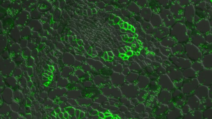
 inoculated with cowpea mosaic virus (CPMV) containing the GFP-gene inserted between the movement protein (MP) and the capsid proteins (CPs) in the viral RNA 2")

