, Tropomyosin (cardiomyocytes, red) and GFP (primordial cardiac layer, green).")
显微镜中的荧光
荧光显微技术是一种特殊的光学显微镜技术。它利用的是荧光色素在一定波长的光激发下发光的能力。通过抗体染色或荧光蛋白标记,可以用这种荧光色素标记感兴趣的蛋白质。这样就可以确定单分子物种的分布、数量及其在细胞内的定位。此外,还可以进行共定位和相互作用研究,使用可逆结合染料(如 Ca2+ 和 fura-2)观察离子浓度,以及观察细胞的内吞和外吞过程。如今,利用荧光显微镜甚至可以对亚分辨率颗粒进行成像。

显微镜测量校准:为什么要校准以及如何校准
显微镜校准可确保用于检测、质量控制 (QC)、故障分析和研发 (R&D) 的测量结果准确一致。本文介绍了校准步骤。使用参照物进行校准可获得可重复的结果,并有助于确保与准则和标准一致。为获得准确一致的结果,建议校准显微镜并定期检查。如有需要,可向校准专家寻求支持。

Visualizing Photoresist Residue and Organic Contamination on Wafers
As the scale of integrated circuits (ICs) on semiconductors passes below 10 nm, efficient detection of organic contamination, like photoresist residue, and defects during wafer inspection is becoming…

Rapidly Visualizing Magnetic Domains in Steel with Kerr Microscopy
The rotation of polarized light after interaction with magnetic domains in a material, known as the Kerr effect, enables the investigation of magnetized samples with Kerr microscopy. It allows rapid…
. With DIC users are able to visualize small height differences on the wafer surface more easily.")
6 英寸晶片检测显微镜,可靠的观察微小高度差
本文介绍了一种 6 英寸晶圆检测显微镜,无论用户的技术水平如何,它都能自动进行可重复的 DIC(微分干涉对比)成像。集成电路 (IC) 芯片和半导体元件的制造需要晶圆检测,以确保不存在影响性能的缺陷。通常使用光学显微镜进行质量控制、故障分析和 R&D 检测。为了有效地观察晶圆上结构之间的微小高度差,可以使用 DIC。
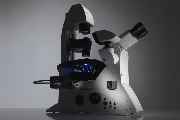
Factors to Consider When Selecting a Research Microscope
An optical microscope is often one of the central devices in a life-science research lab. It can be used for various applications which shed light on many scientific questions. Thereby the…
 and tubulin (magenta), acquired using Viventis Deep. Courtesy of Akanksha Jain, Treutlein Lab ETH-DBSSE Basel (Switzerland).")
如何深入了解类器官和细胞球模型
在本电子书中,您将了解3D细胞培养模型(如类器官和细胞球)成像的关键注意事项。探索创新型显微镜解决方案,来实时记录类器官和细胞球的动态成像过程。

罕见疾病 CRISPR 疗法的开发与风险解除
Fyodor Urnov博士和Sadik Kassim博士最初是在ASGCT 2025会议上作这一按需演讲的,演讲的重点是遗传医学中的一个关键挑战:如何将CRISPR疗法从单一疾病解决方案扩展到平台方法,特别是针对罕见的儿科遗传疾病。Urnov 博士展示了由 Matthew Kan 博士领导的创新基因组研究所的工作,这是 IGI-Danaher Beacon for CRISPR Cures…
 observed with an Ivesta 3 stereo microscope during fly pushing (sorting of the flies). The scale bar length is 1 mm. Image courtesy of M. Benton, EMBL, Heidelberg, Germany.")
Drosophila(果蝇)研究显微镜使用指南
一个多世纪以来,果蝇(典型的黑腹果蝇)一直被用作模式生物。原因之一是果蝇与人类共享许多与疾病相关的基因。果蝇经常被用于发育生物学、遗传学和神经科学的研究。果蝇的优点包括易于饲养且成本低廉、繁殖速度快、基因组完全测序以及可获得各种基因品系。使用徕卡显微镜可以进行高效的果蝇研究。
