
如何选择合适的测量显微镜
使用测量显微镜,用户可以测量样品特征的二维和三维尺寸,这对检测、质量控制、故障分析和研发&D 至关重要。然而,选择合适的显微镜需要评估应用需求以及显微镜的性能、易用性和灵活性。 如今,测量通常以数字方式进行,即使用带有摄像头和软件的显微镜,图像显示在显示器上,而不是通过目镜网线,从而提高了精度和可重复性。使用合适的测量显微镜可靠、快速地分析样品。

显微镜测量校准:为什么要校准以及如何校准
显微镜校准可确保用于检测、质量控制 (QC)、故障分析和研发 (R&D) 的测量结果准确一致。本文介绍了校准步骤。使用参照物进行校准可获得可重复的结果,并有助于确保与准则和标准一致。为获得准确一致的结果,建议校准显微镜并定期检查。如有需要,可向校准专家寻求支持。
 at the edge of a battery electrode acquired with a DVM6 digital microscope.")
电池制造过程中的毛刺检测
毛刺是电池电极片边缘可能出现的缺陷,例如在制造过程中的分切环节。它们可能会因诸如短路等故障导致电池性能下降,并引发安全和可靠性问题。毛刺检测是电池生产质量控制的重要部分,对于生产具有可靠性能和寿命的电池至关重要。通过适当照明的光学显微镜可以在生产过程的关键步骤中快速可靠地对电极上的毛刺进行视觉检测。
 and astrocytes (green) in a cortical spheroid derived from human induced pluripotent stem cells.")
活细胞成像指南
在生命科学各研究领域的广泛应用中,活细胞成像是一种不可或缺的工具,用于观察细胞在尽可能接近活体(即活的、活跃的)状态下的情况。本指南回顾了确保成功进行活细胞成像的各种重要注意事项,并介绍了各种旨在克服常见挑战的高性能解决方案。这些进展使我们能够对细胞生理学和动力学有新的认识。
 and co-stained for nuclear DNA (Hoechst 33342), microtubules (Alexa 555) and F-actin (ATTO 643). Image was captured on Mateo FL.")
用于二维细胞培养的显微镜和AI解决方案
这本电子书探讨了显微镜和AI技术在二维细胞培养工作流程中的整合。报告重点介绍了明视野、相衬和荧光等传统成像方法如何支持常规细胞监测,而 Mateo TL 和 Mateo FL 数字式倒置显微镜则通过自动汇合检查、细胞计数和转染分析提高了可重复性。它还展示了综合数据管理、审计跟踪和样本跟踪如何改进文档和研究的完整性。本书最后展望了未来趋势,包括微流控技术和 2D-3D…
 and tubulin (magenta), acquired using Viventis Deep. Courtesy of Akanksha Jain, Treutlein Lab ETH-DBSSE Basel (Switzerland).")
如何深入了解类器官和细胞球模型
在本电子书中,您将了解3D细胞培养模型(如类器官和细胞球)成像的关键注意事项。探索创新型显微镜解决方案,来实时记录类器官和细胞球的动态成像过程。
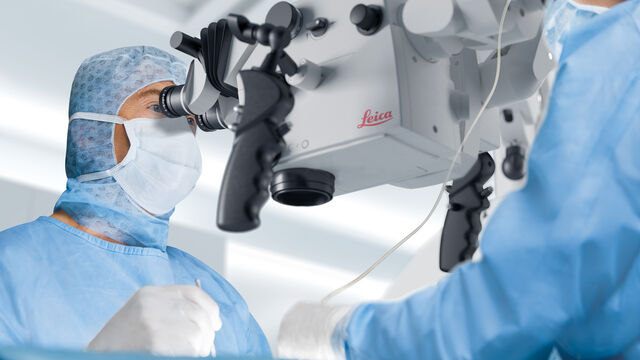
 performing ear, nose and throat (ENT) surgery using the MyVeo surgical visualization headset.")
微血管外科医生的观点:MyVeo 如何实现可视化变革
在这篇文章中,耳鼻喉科医生、头颈部整形外科医生 Andrew T. Huang 博士(医学博士、FACS)分享了使用徕卡微系统公司 MyVeo 头戴显示器进行数字 3D 手术可视化如何改变他的临床实践。对于微血管和神经修复手术,他讨论了如何在手术过程中以舒适放松的姿势帮助自己集中注意力、进行训练并与手术室团队合作。手术可视化显示器还可与手术室无缝集成。了解数字 3D…
 at 2 weeks. Image acquired using Mica.")
如何为深层肌肉组织中的轴突再生成像
这项研究重点介绍了亚伦-李(Aaron Lee)博士对截肢后肌肉移植中神经再生的定位研究。肢体缺失通常会导致生活质量下降,这不仅是因为组织缺失,还因为轴突再生紊乱引起的神经性疼痛。Mica组织学成像和荧光成像可帮助了解神经再生过程中轴突的生长和分支这项研究有助于塑造未来的神经假体接口设计,改善患者的治疗效果。

神经科学研究指南
神经科学通常需要研究具有挑战性的标本,以更好地了解神经系统和疾病。徕卡显微镜帮助神经科学家深入了解神经元功能。
