 which is published by the US FDA (Food & Drug Administration).")
美国联邦法规第21章第11款和其他相关法规简介
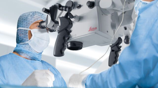
本文概述了在美国(联邦法规第21章第11款)、欧盟(GMP附录11)和中国(NMPA)所用电子记录(数据输入、存储、签名和审批)的法规和指南,这些法规和指南会对医疗器械质量控制的数字化增强检测解决方案产生影响。与纸质记录方法相比,使用显微镜进行数字化增强检测具有更一致和更高效的检测优势。但是,与纸质记录和签名的规定相比,电子记录和签名的规定有明显不同的建议和要求。电子记录的创建、验证、存储和备份应…
Digital Classroom Options
As teachers, you know your big challenge is to catch and keep the students’ attention and the best chance for this is by making the environment interactive. In the case of the Microscopy Classroom, we…
 images")
如何创建EDOF(扩展景深)图像
观看此视频,了解如何使用徕卡显微系统LAS X软件的可选扩展景深(EDOF)功能,快速记录具有较大高度变化样本的清晰光学显微镜图像。以使用徕卡显微镜从低倍到高倍拍摄的电路板EDOF图像为例进行了展示。

页岩和碳酸盐岩的宏观至纳米级孔隙分析
页岩和碳酸盐岩等岩石的物理孔隙度对其储存能力有很大影响。孔隙的几何形状也会影响其渗透率。对可见孔隙空间进行成像,有助于了解物理孔隙空间、孔隙几何形状以及与储存和运输相关的矿物和有机物质阶段。
 stereo microscope for a task like surgery.")
Rodent and Small-Animal Surgery
Learn how you can perform rodent (mouse, rat, hamster) and small-animal surgery efficiently with a microscope for developmental biology and medical research applications by reading this article.

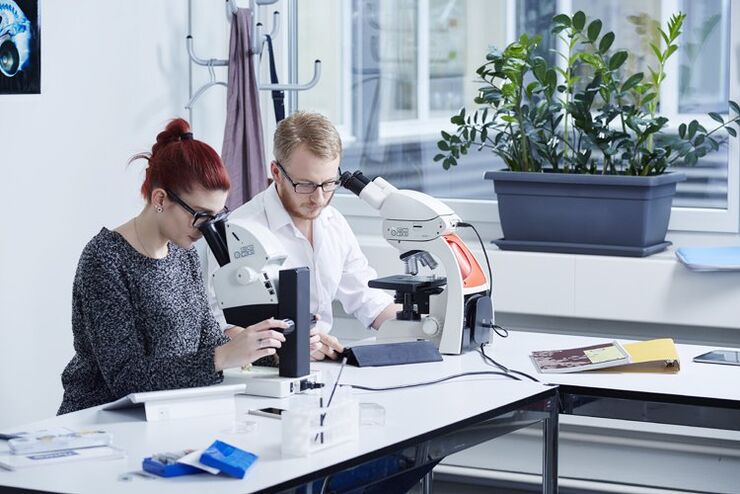
选择学生显微镜需要考虑的因素
对于教师来说,选择教育显微镜并非易事。显微镜必须经得起并非总是小心翼翼的双手的日常使用,必须能够持续运行,还必须符合预算要求。尤其是学生用显微镜,实用性方面起着重要作用: 尺寸、重量、布线和设计在日常使用中非常重要,甚至在决定使用显微镜的设备和附件之前就应考虑到这一点。如果选择得当,教育显微镜将为大中小学的年轻人打开一扇通往微小细节的宇宙之窗,让他们对科学产生足够的兴趣,并将其作为自己的职业。

Gene Editing with CRISPR/Cas9 - Breakthrough in Genome Engineering
The CRISPR/Cas9 system is one of several different bacterial systems for defense against viral attacks. It consists of two main components. One is a small piece of RNA which binds to the viral target…

Imaging and Analyzing Zebrafish, Medaka, and Xenopus
Discover how to image and analyze zebrafish, medaka, and Xenopus frog model organisms efficiently with a microscope for developmental biology applications from this article.

研究果蝇(黑腹果蝇Drosophila melanogaster)
由于每个实验室的需求可能会有很大的差异,本文展示了科学家和技术人员研究果蝇并使用不同显微镜设置的的实例。此外,基于不同果蝇实验室的经验介绍了推荐的工作流程。本文可以作为建立或扩展果蝇实验室时的参考或指南。
