
Loading...
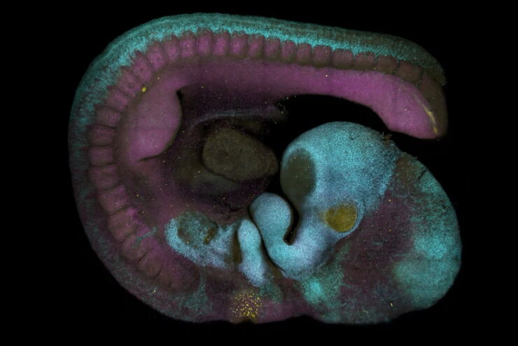
Adding Dimensions to Multiplex Molecular Imaging
Molecular imaging of living specimens offers a means to draw upon the growing body of high-throughput molecular data to better understand the underlying cellular and molecular mechanisms of complex…
Loading...
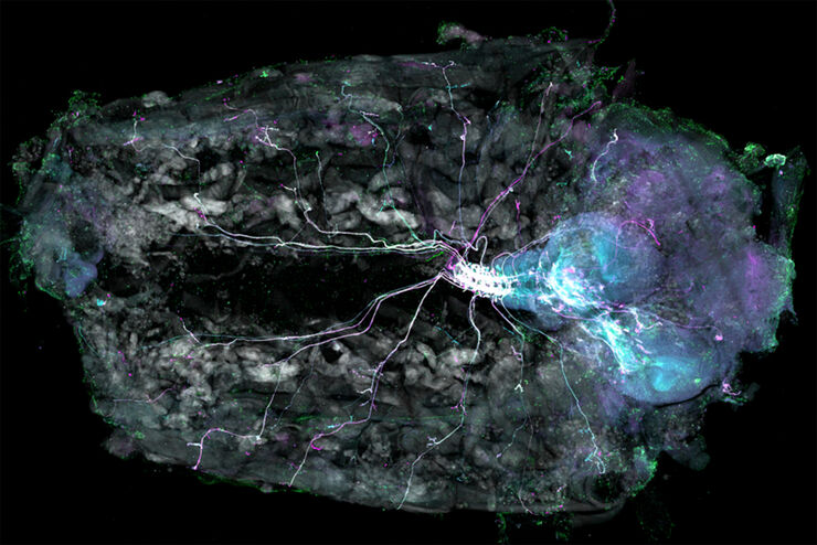
Understanding Motor Sequence Generation Across Spatiotemporal Scales
We have developed a microscopy-based pipeline to characterize a developmentally critical behavior at the pupal stage of development, called the ecdysis sequence. We study brain-wide neuronal activity…
Loading...

Benefits of TauContrast to Image Complex Samples
In this interview, Dr. Timo Zimmermann talks about his experience with the application of TauSense tools and their potential for the investigation of demanding samples such as thick samples or…
Loading...
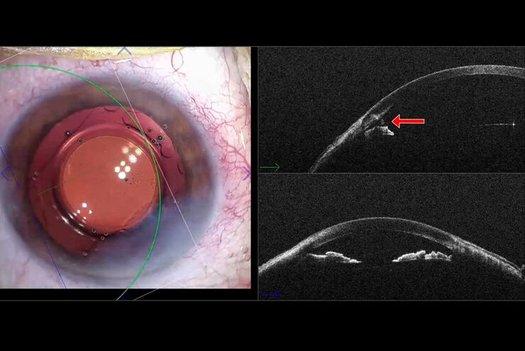
使用EnFocus术中OCT进行白内障切口分析
眼科手术必须满足高精准度的要求,才能让手术操作和组织操作达到理想效果。在帮助眼外科医师在眼科手术中获得高精准度和提升手术效果方面,眼科手术显微镜发挥着重要的作用。
Loading...
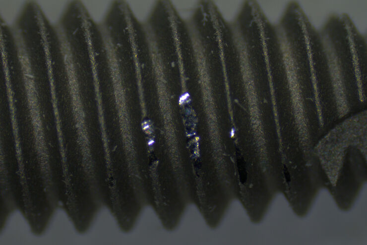
对医疗器械进行手动目视检查为何如此之难?
本文旨在对医疗器械行业中普遍存在的手动目视检查是如何导致不一致结果这一问题进行讨论。此外,本文还讨论了质量经理和操作员在进行手动检查时所面临的挑战,以及他们是如何使用数字化增强检查解决方案来克服这些挑战的。
Loading...

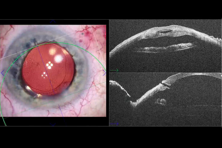
术中光学相干断层扫描(OCT)辅助下增生性玻璃体视网膜病变的手术治疗
对患者及其外科医生而言,增生性玻璃体视网膜病变(PVR)是近期孔源性视网膜脱离(RD)后的一大麻烦。尽管初始手术效果极佳,但PVR是导致RD复发的最常见原因,通常导致患者最终视力下降。即使患者最初未发生黄斑脱离,在既往玻璃体切除术后视网膜可再次快速脱离,常累及黄斑,可能需要多次手术来保持视网膜永久附着。
Loading...
Fast, High-quality Vitrification with the EM ICE High Pressure Freezer
The EM ICE High Pressure Freezer was developed with a unique freezing principle and uses only a single pressurization and cooling liquid: liquified nitrogen (LN2). This design enables three major…
Loading...
![[Translate to chinese:] Cryo FIB lamella - Overlay of SEM and confocal fluorescence image. Target structure in yeast cells (nuclear pore proteine Nup159-Atg8-split Venus, red) marked by an arrow. Scale bar: 5 µm. Alegretti et al., Nature 586, 796-800 (2020).](/fileadmin/_processed_/c/d/csm_Targeting_Nuclear_Pore_Complexes_teaser_16478dc18a.jpg "[Translate to chinese:] Cryo FIB lamella - Overlay of SEM and confocal fluorescence image")
使用冷冻共聚焦显微镜定位活性循环核孔复合物
本文介绍了如何利用冷冻光学显微镜,尤其是冷冻共焦显微镜来提高冷冻工作流程的可靠性。评估了EM网格和样品的质量,并分析了目标结构的分布。本文展示了如何将冷冻共焦3D数据投射到SEM图像上,将感兴趣结构可靠地保留在FIB切割的薄片内,以便在冷冻TEM中进行进一步研究。