
Loading...

Overcoming Observational Challenges in Organoid 3D Cell Culture
Learn how to overcome challenges in observing organoid growth. Read this article and discover new solutions for real-time monitoring which do not disturb the 3D structure of the organoids over time.
Loading...

How to Streamline Your Histology Workflows
Streamline your histology workflows. The unique Fluosync detection method embedded into Mica enables high-res RGB color imaging in one shot.
Loading...
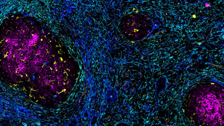
Accelerating Discovery for Multiplexed Imaging of Diverse Tissues
Explore IBEX: Open-source multiplexed imaging. Join the collaborative IBEX Imaging Community for optimized tissue processing, antibody selection, and human atlas construction.
Loading...

Notable AI-based Solutions for Phenotypic Drug Screening
Learn about notable optical microscope solutions for phenotypic drug screening using 3D-cell culture, both planning and execution, from this free, on-demand webinar.
![[Translate to chinese:] Hepatocellular Carcinoma with 13 biomarkers shown – Beta-Catenin, CD3D, CD4, CD8a, CD31, CD44, CD163, DAPI, PanCK, PCK26, PD1, SMA, and Vimentin.](/fileadmin/_processed_/5/b/csm_Hepatocellular_Carcinoma_13_Markers_Zoom2_bc27c21cf2.jpg "[Translate to chinese:] Hepatocellular Carcinoma with 13 biomarkers shown – Beta-Catenin, CD3D, CD4, CD8a, CD31, CD44, CD163, DAPI, PanCK, PCK26, PD1, SMA, and Vimentin.")
Loading...
![[Translate to chinese:] Co-detection of 10 extracellular matrix proteins and 3 topographical tissue landmarks by multiplex immunostaining within a single high-grade fibrous hotspot from a human hepatocellular carcinoma](/fileadmin/_processed_/0/8/csm_Single_high-grade_fibrous_hotspot_from_a_human_hepatocellular_carcinoma_e5541282bd.jpg "[Translate to chinese:] Co-detection of 10 extracellular matrix proteins and 3 topographical tissue landmarks by multiplex immunostaining within a single high-grade fibrous hotspot from a human hepatocellular carcinoma")
肝细胞癌中癌症干细胞位点的原位鉴定
在这里,我们探索了一种突破性的多重免疫检测方法,通过多重成像对细胞外基质(ECM)特征进行原位定位,从而识别肝细胞癌(HCC)内的癌症干细胞龛。
Loading...
![[Translate to chinese:] Pancreatic Ductal Adenocarcinoma with 11 Apoptosis biomarkers shown – BAK, BAX, BCL2, BCLXL, Caspase9, CIAP1, NaKATPase, PCK26, SMAC, Vimentin, and XIAP.](/fileadmin/academy/2023/Gated_content/Pancreatic_Ductal_Adenocarcinoma_11_Apoptosis_Markers_ROI5.jpg "[Translate to chinese:] Pancreatic Ductal Adenocarcinoma with 11 Apoptosis biomarkers shown – BAK, BAX, BCL2, BCLXL, Caspase9, CIAP1, NaKATPase, PCK26, SMAC, Vimentin, and XIAP.")
与卢克-加蒙(Luke Gammon)一起多重成像:推进您的空间生物学研究
多重成像是一种功能强大的技术,可让研究人员同时观察单个样本中的多个目标。这对于研究复杂的生物系统尤为重要,可以帮助研究人员更好地了解不同分子和途径之间是如何相互作用的。
Loading...

如何使用数字化显微镜测定细胞汇合度
本文介绍了如何以一致性的方式测量细胞汇合度。对于生命科学研究领域,例如癌症生物学、干细胞或再生医学,研究通常需要特定生长条件下的细胞。这些条件包括定期检查的细胞形态和汇合度。
Loading...

如何对细胞培养进行快速正确的检测
本文介绍了对培养贴壁细胞系进行传代的一般工作流程及步骤说明。哺乳类细胞体外培养是在癌症、药物开发、组织工程、干细胞、疾病细胞和分子生物学等生物医学研究领域进行临床和药物研究的重要模型。要想成功维持细胞系,需要通过控制生长条件来维持细胞生理和表型的稳定性。定期监测细胞生长,对细胞进行传代培养以确保连续性。