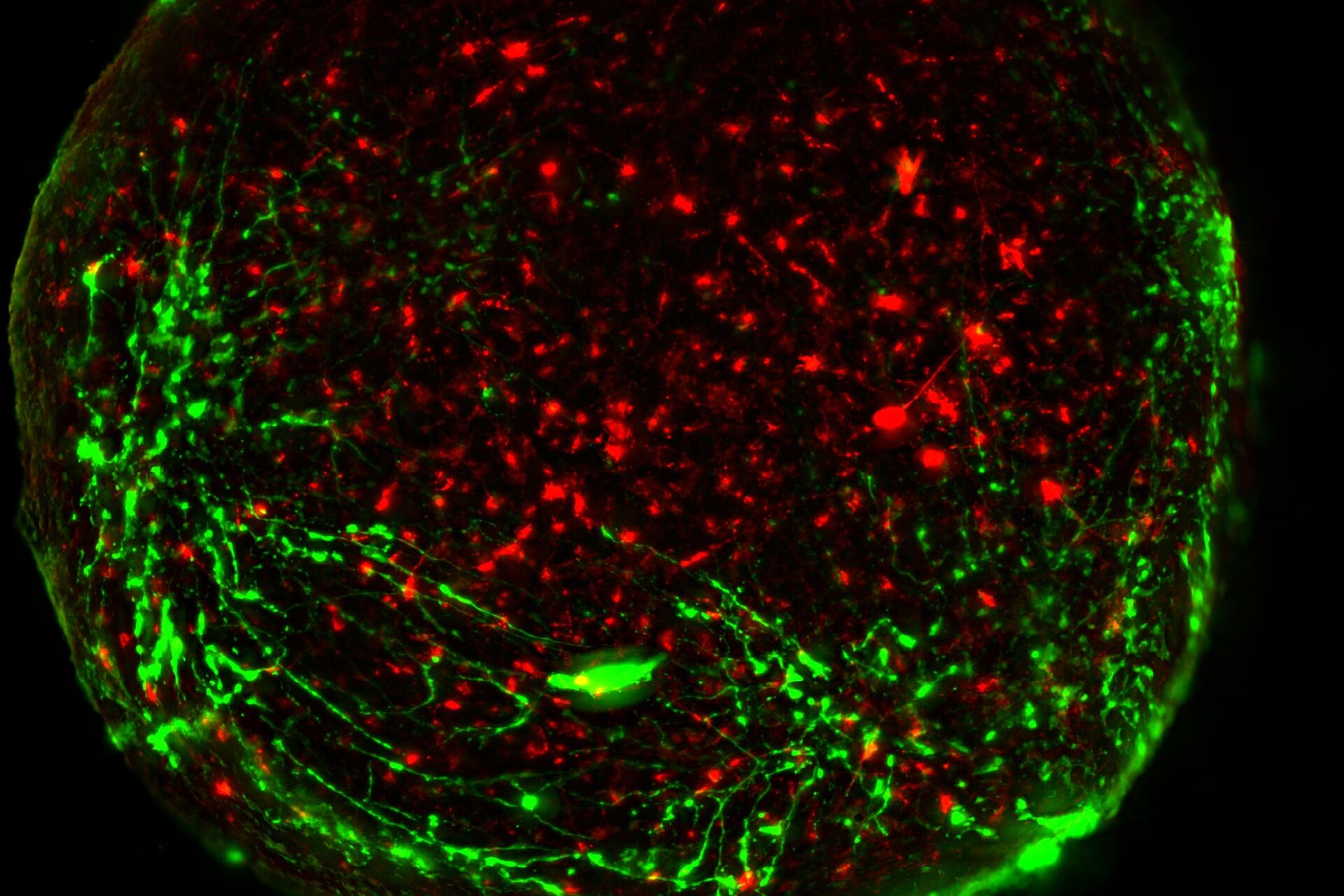
活细胞成像

使用现在已开发的各种荧光蛋白和多色探针几乎可以标记任何分子。 对囊泡、细胞器、细胞和组织中的蛋白质动力学成像的能力为了解细胞在健康和疾病状态下如何工作提供了新的洞察力。 这些包括有丝分裂、胚胎发育和细胞骨架变化等过程的时空动态。

研究活细胞时,常见的障碍包括光毒性和光损伤。 要捕捉快速的生物过程,关键是保持细胞健康并获得清晰的图像,确保数据可靠、无伪影。 活‐细胞显微成像通常需要在图像质量与细胞健康之间作出取舍。 在成像过程中必须保持特定的环境条件,以免细胞发生变化。

各种高性能的徕卡成像解决方案可以克服活细胞成像的这些挑战,有助发现细胞生理学和动力学方面的新信息。

立即联系!

我们的活细胞成像应用解决方案专家将竭诚为您提供建议.

您的活细胞成像需求

要想成功地进行活细胞成像实验,使用合适的平台至关重要。 在选择用于活细胞成像的光学显微镜时,应考虑以下 3 个变量:检测器灵敏度(信噪比)、样本活性和图像采集速度。 适合活细胞应用的方法能够在不损伤细胞的情况下对动态事件成像,因为细胞损伤会影响结果。 活细胞成像主要使用荧光显微镜进行。

徕卡显微系统的 THUNDER 成像系统、一款 Microhub 的 Mica、STELLARIS 共聚焦平台和 FLIM 提供了最新的宽场和共聚焦成像创新技术,可快速进行 3D 活细胞成像。

宽场显微镜可灵活激发和快速采集,通常用于对细胞动态和发育进行长时间成像。 共聚焦显微镜通常用于研究亚细胞动态事件。 多光子显微镜可使用较长波长的光激发,可减少光漂白并延长细胞活性。 最后,荧光寿命成像 (FLIM) 可用于研究细胞中的快速动态信号事件。

徕卡DMi8倒置显微镜 DMi8 S 高速成像平台

模块化的 DMi8 倒置显微镜是 DMi8 S 平台的核心。DMi8 S 平台是适用于日常活细胞研究的完整解决方案。不管是精确跟踪培养皿中单个细胞的发育,筛选多个分析,获取单分子级的清晰度,还是梳理复杂过程的行为,DMi8 S 系统都能让您看得更多、看得更快,让您发现隐藏的信息。

成像过程中的细胞活性和动力学

徕卡显微系统为您提供活细胞成像方面的智能创新。 我们的解决方案可帮助您获得最佳的图像质量,同时保护好您的样本。 大多数细胞过程在三维空间中随着时间的推移进行。 因此,若要掌握全面的情况,必须以四个维度(XYZ 和时间)对细胞成像。 延时成像方法可捕捉从几秒到几个月内的细胞事件。 也可以在特定时间点对细胞重复成像。 为了在这个过程中保护细胞活性,活细胞成像时需要温度、酸碱度和湿度都受到控制。 曝光量也应在最低限度,以免发生光毒性。

徕卡显微系统提供的成像解决方案有助于优化您对活细胞的研究,即使是长时间的研究也同样如此。 它们能提供必要的图像对比度和分辨率,有助动态事件的分析。 有些徕卡系统还可以实现高速成像,可以避免相同时间点的标签之间发生时空失配,因此不会错过任何关键的细胞事件。

活细胞温和成像

使用STELLARIS,您不必再为了获得出色的图像而损伤样本健康。 无论您是需要很长的采集时间进行高分辨率的3D图像重构,还是需要高帧频来捕捉高速动态事件,我们的高效信号探测技术可确保您在防止样本受到光漂白和光毒性影响的前提下完成所需要的实验。

相关文章

阅读我们的最新文章

作为徕卡显微系统有限公司 的信息门户,ScienceLab (徕卡课堂) 提供许多关于显微技术主题的科研和教学材料。其内容宗旨在于为初学者、有经验的从业者和科学家等的日常工作和实验提供支持。

更多文章
Mouse cortical neurons. Transgenic GFP (green). Image courtesy of Prof. Hui Guo, School of Life Sciences, Central South University, China

How Microscopy Helps the Study of Mechanoceptive and Synaptic Pathways

In this podcast, Dr Langenhan explains how microscopy helps his team to study mechanoceptive and synaptic pathways, their challenges, and how they overcome them.
Phase-contrast image of a MDCK-cell culture and its respective confluency measured by the Mateo TL microscope.

如何使用数字化显微镜测定细胞汇合度

本文介绍了如何以一致性的方式测量细胞汇合度。对于生命科学研究领域,例如癌症生物学、干细胞或再生医学,研究通常需要特定生长条件下的细胞。这些条件包括定期检查的细胞形态和汇合度。
Combining spectrally resolved detection and fluorescence lifetime multiplexing

Live-Cell Fluorescence Lifetime Multiplexing Using Organic Fluorophores

On-demand video: Imaging more subcellular targets by using fluorescence lifetime multiplexing combined with spectrally resolved detection.
Living HeLa cells stained with WGA-488 (yellow), SPY-Actin (cyan), and SiR-Tubulin (magenta). Instant Computational Clearing (ICC) was applied.

How to Perform Dynamic Multicolor Time-Lapse Imaging

Live-cell imaging sheds light on diverse cellular events. As many of these events have fast dynamics, the microscope imaging system must be fast enough to record every detail. One major advantage of…
MDCK cysts on day 9

How To Perform Fast & Stable Multicolor Live-Cell Imaging

With the help of live-cell imaging researchers gain insights into dynamic processes of living cells up to whole organisms. This includes intracellular as well as intercellular activities. Protein or…
Wt1b-positive myocardial cell adopting an epicardial cell morphology.

Wt1 Genes Can Induce a Cardiomyocyte to Epicardial-like Cell Fate Transition

From this study, it was concluded that Wt1 plays a yet undescribed role for cardiomyocyte differentiation by repressing chromatin opening at specific genomic loci and that sustained ectopic expression…
Multi-color SRS image of a tri-cellular cancer spheroid

The Potential of Coherent Raman Scattering Microscopy at a Glance

Coherent Raman scattering microscopy (CRS) is a powerful approach for label-free, chemically specific imaging. It is based on the characteristic intrinsic vibrational contrast of molecules in the…

A Versatile Palette of Fluorescent Probes

Researchers at the Max Planck Institute for Medical Research in Heidelberg have developed a general strategy to synthesize live-cell compatible fluorogenic probes, and the result are the new MaP (Max…
3D-volume-rendered light-sheet microscope image of a spheroid showing depth coding in different colors.

Imaging of Anti-Cancer Drug Uptake in Spheroids using DLS

Spheroid 3D cell culture models mimic the physiology and functions of living tissues making them a useful tool to study tumor morphology and screen anti-cancer drugs. The drug AZD2014 is a recognized…
Spontaneous colon adenoma

Intravital Microscopy of Cancer

Join our guest speaker Prof Dr Jacco van Rheenen, as he presents his work on the identity, behavior and fate of cells that drive the initiation and progression of cancer.

关于活细胞成像

除了细胞或器官的结构组织,细胞动态过程是一个功能生物实体的主要贡献者。当然,这些过程可以在活细胞中通过非侵入性技术如光学方法观察到,统称为“活细胞成像”方法。活细胞成像涵盖了所有用显微镜观察活细胞的技术——从用体视显微镜观察胚胎发生,到用复合显微镜研究细胞生长,直到用荧光染料或荧光蛋白研究细胞的生理状态或细胞运输。尽管对实验人员和设备(如成像系统,温度、CO2浓度控制)都要求很高,但活细胞成像技术提供的结果是当今研究不可或缺的。

有兴趣了解更多?

请与我们的专家交流。 我们很乐意回答您的所有问题和疑虑。

联系我们

您想获取专人咨询吗? Show local contacts

Scroll to top