
![[Translate to chinese:] Virally labeled neurons (red) and astrocytes (green) in a cortical spheroid derived from human induced pluripotent stem cells. THUNDER Model Organism Imagerwith a 2x 0.15 NA objective at 3.4x zoomwas used to produce this 425 μm Z-stack (26 positions), which is presented here as an Extended Depth of Field(EDoF)projection.](/fileadmin/_processed_/e/c/csm_THUNDER_Imager_Model-Org_Header-Gallery-Neuroscience_4652e132e6.jpg "[Translate to chinese:] Virally labeled neurons (red) and astrocytes (green) in a cortical spheroid derived from human induced pluripotent stem cells.")
Loading...
多彩图库
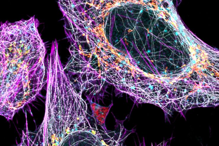
荧光多色显微技术是多重成像技术的一个方面,可在同一实验中观察和分析同一样本中的多种元素--每种元素都标记有不同的荧光染料。这不仅能提高实验效率,还能获得更可靠、更有意义的结果,从而了解细胞和组织内的复杂过程。本图集展示了使用THUNDER和STELLARIS平台获得的标有多种荧光探针的样本图像。
Loading...
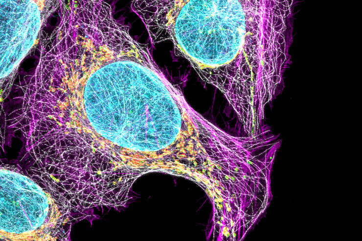
细胞生物学图片库
细胞生物学研究细胞的结构、功能和行为,包括细胞新陈代谢、细胞周期和细胞信号传导。荧光显微镜是细胞生物学家工具包中不可或缺的一部分。宽场显微镜和共聚焦显微镜被广泛用于观察细胞内复杂结构的细节。
Loading...
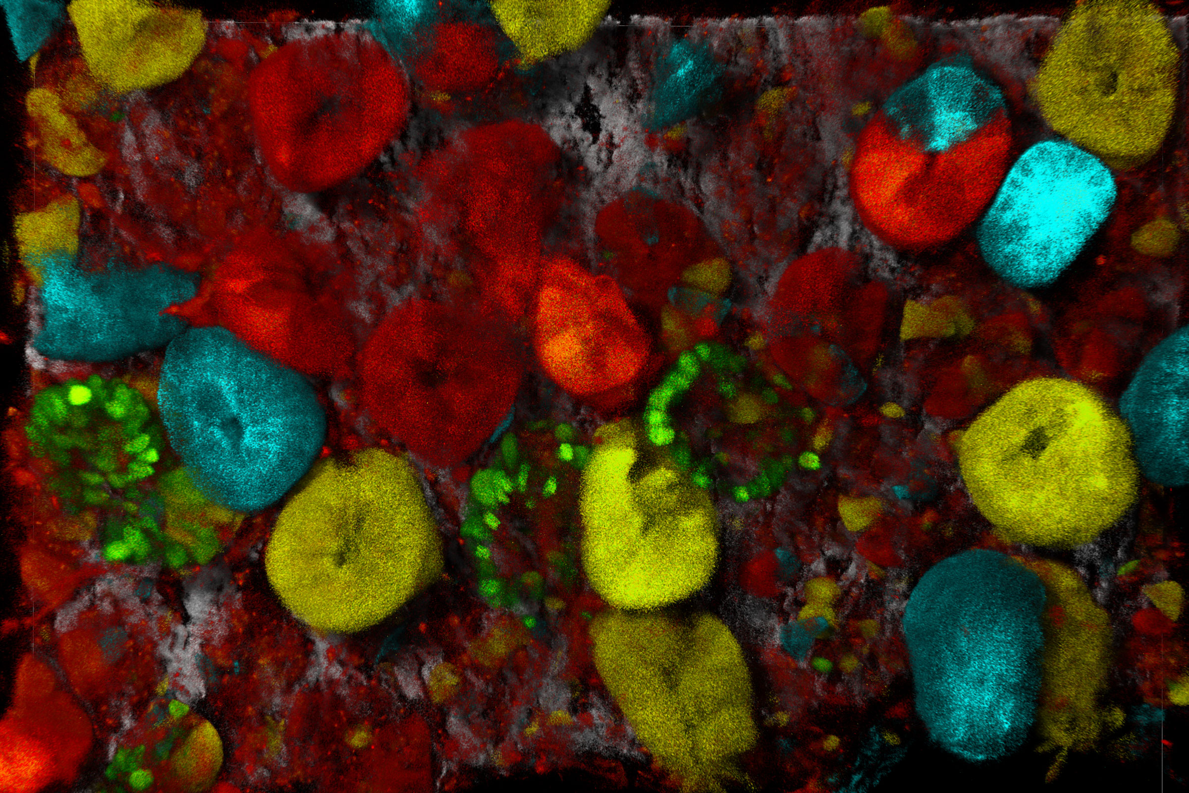
Dissecting Proteomic Heterogeneity of the Tumor Microenvironment
This lecture will highlight cutting edge applications in applying laser microdissection and microscaled quantitative proteomics and phosphoproteomics to uncover exquisite intra- and inter-tumor…
Loading...

Developmental Biology Image Gallery
Developmental biology explores the development of complex organisms from the embryo to adulthood to understand in detail the origins of disease. This category of the gallery shows images about…
Loading...
Kinetochore Assembly during Mitosis with TauSTED on 3D
Three-dimensional organization of the mitotic spindle together with the distribution of CENP-C and BUB1 based on TauSTED with multiple STED lines (592, 660 and 775 nm) can provide insights on…
Loading...
How to Quantify Changes in the Metabolic Status of Single Cells
Metabolic imaging based on fluorescence lifetime provides insights into the metabolic dynamics of cells, but its use has been limited as expertise in advanced microscopy techniques was needed.
Now,…
Loading...
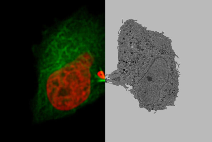
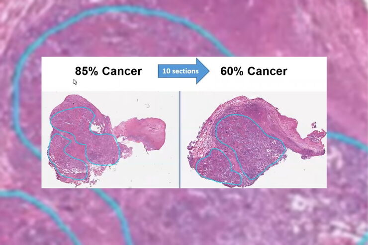
将动态活细胞数据融入超微结构
采用徕卡Nano的工作流程,可以避免过去如海底捞针似的寻找。利用光电关联显微技术,在适当的时间直接鉴别出正确的细胞,并将动态的活细胞数据融入其超微结构中。