Loading...
![[Translate to chinese:] 40x magnification of organoids cluster taken on Mateo TL.Cell type: esophageal squamous carcinoma; scale bar 15µm. Courtesy of bioGenous, China. [Translate to chinese:] 40x magnification of organoids cluster taken on Mateo TL.Cell type: esophageal squamous carcinoma; scale bar 15µm. Courtesy of bioGenous, China.](/fileadmin/_processed_/c/a/csm_Esophageal_squamous_carcinoma_scale__bar_15_m_7bfa184294.jpg)
克服类器官三维细胞培养中的观察挑战
类器官在细胞生物学和药物发现中至关重要,因为它们能够模拟体内细胞的复杂性和结构,有助于癌症等微环境至关重要的疾病研究。类器官可根据患者的基因型进行定制,这也有助于个性化医学研究。
Loading...

Notable AI-based Solutions for Phenotypic Drug Screening
Learn about notable optical microscope solutions for phenotypic drug screening using 3D-cell culture, both planning and execution, from this free, on-demand webinar.
Loading...
![[Translate to chinese:] The principle of the FusionOptics technology [Translate to chinese:] The principle of the FusionOptics technology: Of the two separate beam paths (1), one provides depth of field (2) and the other high resolution (3). In the brain, the two images of the sample are merged into a single, optimal 3D image (4).](/fileadmin/_processed_/3/c/csm_csm_fusionoptics_pcb-inspection_e71950a5dd_271afc1b85.jpg)
FusionOptics融合光学(徕卡专利技术)高分辨率和大景深结合
人类对视觉环境的感受,80%通过视觉感知获得。如果没有空间视觉,我们几乎无法确定方向。我们的视觉皮质和大脑皮层通过复杂的过程,巧妙地处理着从眼睛看到的图像信号。最近几十年,神经科学已经大量了解该过程。故由徕卡显微系统、苏黎世大学神经信息学研究所和瑞士联邦理工学院共同开展的一项研究,显示了我们的大脑如何灵活有效地结合视觉信号创造出最佳空间图像。研究结果为立体显微技术创新奠定了基础,该创新从分辨率和聚…
Loading...
![[Translate to chinese:] Branched organoid growing in collagen where the Nuclei are labeled blue. To detect the mechanosignaling process, the YAP1 is labeled green. [Translate to chinese:] Branched organoid growing in collagen where the Nuclei are labeled blue. To detect the mechanosignaling process, the YAP1 is labeled green.](/fileadmin/_processed_/a/e/csm_Branched_organoid_growing_in_collagen_083254eb44.jpg)
检查癌症类器官的发展进程
德国慕尼黑工业大学的Andreas Bausch实验室研究细胞和生物体中不同结构和功能形成的细胞和生物物理机制。他的团队设计了新的策略、方法和分析工具,以量化微米和纳米等级的发展机制和动态过程。关键研究领域包括干细胞和类器官,从乳腺类器官到胰腺癌类器官,以更好地了解疾病模型。
Loading...
![[Translate to chinese:] THY1-EGFP labeled neurons in mouse brain processed using the PEGASOS 2 tissue clearing method. Neurons were traced using Aivia’s 3D Neuron Analysis – FL recipe. Image credit: Hu Zhao, Chinese Institute for Brain Research. [Translate to chinese:] THY1-EGFP labeled neurons in mouse brain processed using the PEGASOS 2 tissue clearing method, imaged on a Leica confocal microscope. Neurons were traced using Aivia’s 3D Neuron Analysis – FL recipe. Image credit: Hu Zhao, Chinese Institute for Brain Research.](/fileadmin/_processed_/f/1/csm_Neurons_in_mouse_brain_with_Aivia_3D_Neuron_Analysis_9aa755e1c5.jpg)
借助人工智能,揭示复杂而密集的神经元图像中的洞察
神经元的3D形态学分析通常需要使用不同的成像模式,捕捉多种类型的神经元,并在各种密度下相连的传统Leica SP8显微镜采集多达解神经元的形态,这对许多研究人员来说仍然是一个耗时的挑战。
Loading...

What are the Challenges in Neuroscience Microscopy?
eBook outlining the visualization of the nervous system using different types of microscopy techniques and methods to address questions in neuroscience.

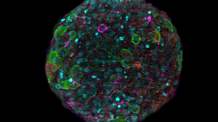
![[Translate to chinese:] Murine esophageal organoids (DAPI, Integrin26-AF 488, SOX2-AF568) imaged with the THUNDER Imager 3D Cell Culture. Courtesy of Dr. F.T. Arroso Martins, Tamere University, Finland. [Translate to chinese:] Murine esophageal organoids (DAPI, Integrin26-AF 488, SOX2-AF568) imaged with the THUNDER Imager 3D Cell Culture. Courtesy of Dr. F.T. Arroso Martins, Tamere University, Finland.](/fileadmin/_processed_/f/f/csm_THUNDER_Imager_3D_Cell_Culture_Murine-esophageal-organoid_LVCC_9bc587500f.jpg)
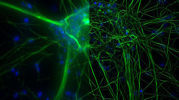
![[Translate to chinese:] Mouse cortical neurons. Transgenic GFP (green). Image courtesy of Prof. Hui Guo, School of Life Sciences, Central South University, China [Translate to chinese:] Mouse cortical neurons. Transgenic GFP (green). Image courtesy of Prof. Hui Guo, School of Life Sciences, Central South University, China](/fileadmin/_processed_/2/a/csm_THUNDER_Imager_Mouse_cortical_neuron_9d4054774d.jpg)
![[Translate to chinese:] Cancer cells [Translate to chinese:] Cancer cells](/fileadmin/_processed_/6/b/csm_Cancer_cells_0188d06359.jpg)