
![[Translate to chinese:] Images of the same area of a processed wafer taken with standard (left) and oblique (right) brightfield illumination using a Leica compound microscope. The defect on the wafer surface is clearly more visible with oblique illumination.](/fileadmin/_processed_/9/1/csm_Processed_wafer_standard_and_oblique_brightfield_illumination_fb296b5b5d.jpg "[Translate to chinese:] Images of the same area of a processed wafer taken with standard (left) and oblique (right) brightfield illumination using a Leica compound microscope. The defect on the wafer surface is clearly more visible with oblique illuminati")
Loading...
![[Translate to chinese:] Intensity distribution (arbitrary color coding) of an image of two points where the distance between them corresponds to the Rayleigh criterion.](/fileadmin/_processed_/3/1/csm_Intensity_distribution_Rayleigh_criterion_58ccd036cd.jpg "[Translate to chinese:] Intensity distribution (arbitrary color coding) of an image of two points where the distance between them corresponds to the Rayleigh criterion.")
显微镜分辨率:概念、因素和计算
在显微镜学中,‘分辨率’一词用于阐述显微镜对细节进行区分的能力。换言之,这是样本内两个能被观察人员或者显微镜摄像头区分的实体点之间的最小距离。显微镜的分辨率本质上与光学元件的数值孔径(NA)以及用于观察样本标本的光波长有关。此外,我们必须考虑Ernst Abbe于1873年首次提出的衍射极限。本文章包含了这些概念的历史介绍并使用相对简单的术语对其进行了解释。
Loading...
")
A Brief History of Light Microscopy – From the Medieval Reading Stone to Super-Resolution
The history of microscopy begins in the Middle Ages. As far back as the 11th century, plano-convex lenses made of polished beryl were used in the Arab world as reading stones to magnify manuscripts.…
Loading...

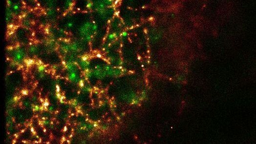
Total Internal Reflection Fluorescence (TIRF) Microscopy
Total internal reflection fluorescence (TIRF) is a special technique in fluorescence microscopy developed by Daniel Axelrod at the University of Michigan, Ann Arbor in the early 1980s. TIRF microscopy…
Loading...

Applications of TIRF Microscopy in Life Science Research
The special feature of TIRF microscopy is the employment of an evanescent field for fluorophore excitation. Unlike standard widefield fluorescence illumination procedures with arc lamps, LEDs or…
Loading...
Super-Resolution GSDIM Microscopy
The nanoscopic technique GSDIM (ground state depletion microscopy followed by individual molecule return) provides a detailed image of the spatial arrangement of proteins and other biomolecules within…