Loading...
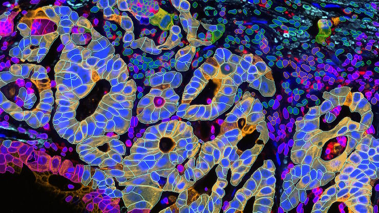
Transforming Multiplexed 2D Data into Spatial Insights Guided by AI
Aivia 13 handles large 2D images and enables researchers to obtain deep insights into microenvironment surrounding their phenotypes with millions of detected objects and automatic clustering up to 30…
Loading...
![[Translate to chinese:] Single cell datasets [Translate to chinese:] Single cell datasets](/fileadmin/_processed_/8/8/csm_Single_cell_datasets_SPARCS_441a58445d.jpg)
利用 SPARCS 探索亚细胞空间表型
功能日益强大的显微镜可提供信息丰富的各种细胞表型数据。如果与深度学习的最新进展相结合,这将成为在基因筛选中读出感兴趣的生物表型的理想技术。在本网络讲座中,您将了解到空间分辨 CRISPR 筛选 (SPARCS),这是一种利用自动化高速激光显微切割技术在人类基因组尺度上揭示各种亚细胞空间表型的平台。
Loading...
![[Translate to chinese:] THY1-EGFP labeled neurons in mouse brain processed using the PEGASOS 2 tissue clearing method. Neurons were traced using Aivia’s 3D Neuron Analysis – FL recipe. Image credit: Hu Zhao, Chinese Institute for Brain Research. [Translate to chinese:] THY1-EGFP labeled neurons in mouse brain processed using the PEGASOS 2 tissue clearing method, imaged on a Leica confocal microscope. Neurons were traced using Aivia’s 3D Neuron Analysis – FL recipe. Image credit: Hu Zhao, Chinese Institute for Brain Research.](/fileadmin/_processed_/f/1/csm_Neurons_in_mouse_brain_with_Aivia_3D_Neuron_Analysis_9aa755e1c5.jpg)
借助人工智能,揭示复杂而密集的神经元图像中的洞察
神经元的3D形态学分析通常需要使用不同的成像模式,捕捉多种类型的神经元,并在各种密度下相连的传统Leica SP8显微镜采集多达解神经元的形态,这对许多研究人员来说仍然是一个耗时的挑战。
Loading...
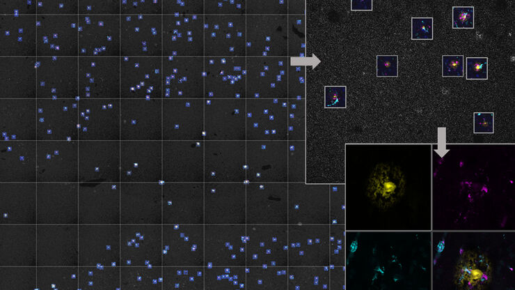
人工智能显微成像能够高效检测稀有事件
对稀有事件进行定位和选择性成像是许多生物样本研究过程的关键。然而,由于时间限制和高度的复杂性,有些实验无法做到,从而限制了获得新发现的前景。通过基于人工智能的显微成像检测稀有事件,这种工作流程将智能样本导航、图像采集工具和人工智能驱动的图像分析等不同功能融合起来共同协作,能够克服上述局限性。
Loading...
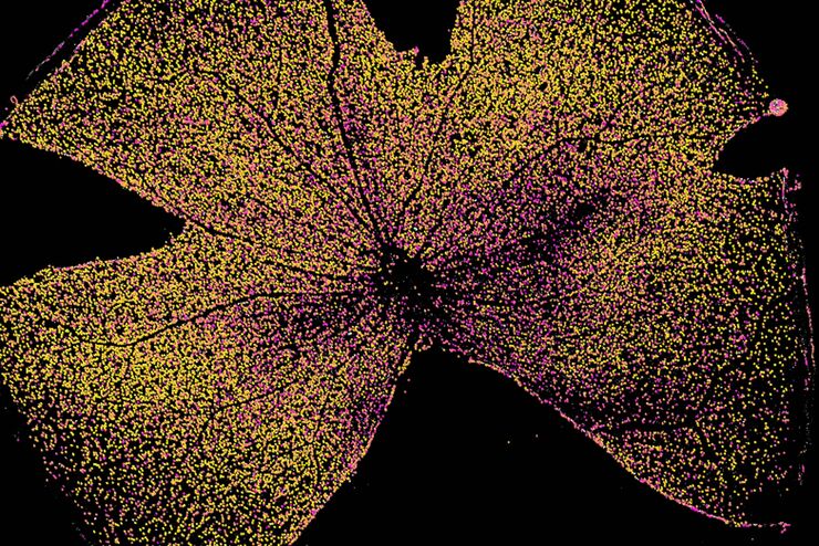
快速、高灵敏度成像和人工智能辅助分析
The specificity of fluorescence microscopy allows researchers to accurately observe and analyze biological processes and structures quickly and easily, even when using thick or large samples. However,…
Loading...
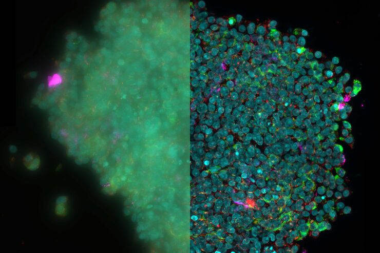
为活细胞成像创造新选择
对厚实的活体样本进行成像时,主要挑战之一是获得图像质量与组织完整性之间的平衡。长时间的图像采集期间,弱信号光会导致低信号水平,导致图像对比度低以及分割和分析困难。需要通过高剂量成像或高时间分辨率成像技术加强信号强度时,这一问题更加突出。一个常见问题是:我如果快速成像、一次完成,会不会造成样本过度漂白或者细胞死亡?
Loading...
![[Translate to chinese:] Image of fixed U2OS cell expressing mEmerald-Tomm20 denoised using a 3D RCAN model trained with matching low and high SNR image pairs acquired on an iSIM system. [Translate to chinese:] Image of fixed U2OS cell expressing mEmerald-Tomm20 denoised using a 3D RCAN model trained with matching low and high SNR image pairs acquired on an iSIM system.](/fileadmin/_processed_/8/7/csm_Fixed_U2OS_cell_expressing_mEmerald-Tomm20_Teaser_01c47585d7.jpg)
人工智能显微图像分析-介绍
人工智能引领的显微图像分析和可视化是用于数据驱动型科学发现的一项强大工具。人工智能技术可以帮助研究人员应对具有挑战性的成像应用,让他们能够从图像中获取更多的信息。
Loading...
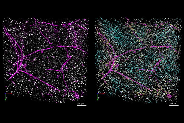
精确分析宽视野荧光图像
利用荧光显微镜的特异性,即便是使用厚样品和大尺寸样品,研究人员也能够快速轻松地准确观察和分析生物学过程和结构。然而,离焦荧光会提高背景荧光,降低对比度,影响图像的精确分割。THUNDER 与Aivia 的组合可以有效解决这一问题。前者可以消除图像模糊,后者会使用人工智能技术自动分析宽视野图像,提高操作速度和精确性。下面,我们来详细了解下这一协作方法。

![[Translate to chinese:] How is microscopy used in spatial biology? A microscopy guide. [Translate to chinese:] How is microscopy used in spatial biology - Teaserimage](/fileadmin/_processed_/0/f/csm_spatial-biology-ebook-tease_5ac6218fa3.jpg)