
Loading...
![[Translate to chinese:] Multicolor TauSTED Xtend 775 for Cell Biology applications that require nanoscopy resolution for multiple cellular components. Cells showing vimentin fibrils (AF 594), actin network (ATTO 647N), and nuclear pore basket (CF 680R). Sample courtesy of Brigitte Bergner, Mariano Gonzales Pisfil, Steffen Dietzel, Core Facility Bioimaging, Biomedical Center, Ludwig-Maximilians-University, Munich, Germany.](/fileadmin/_processed_/5/4/csm_Triple_color_fixed_sample_TauSTED_Xtend_2_3_5d4d9caa21.jpg "[Translate to chinese:] Multicolor TauSTED Xtend 775 for Cell Biology applications that require nanoscopy resolution for multiple cellular components. Cells showing vimentin fibrils (AF 594), actin network (ATTO 647N), and nuclear pore basket (CF 680R).")
STED样品制备指南
这份指南旨在帮助用户优化受激发射损耗(STED)纳米成像的样品制备,特别是在使用徕卡微系统的STED显微镜时。它提供了单色STED成像用荧光标记的概述,并对其性能进行了评级。
Loading...

Epi-Illumination Fluorescence and Reflection-Contrast Microscopy
This article discusses the development of epi-illumination and reflection contrast for fluorescence microscopy concerning life-science applications. Much was done by the Ploem research group…
Loading...
如何为免疫荧光显微镜制备样本
免疫荧光(IF)是一种用于可视化观察细胞内过程、状态和结构的强大工具。IF制剂可通过多种显微镜技术(如激光共聚焦、宽场荧光、全内反射成像等)来加以分析,具体取决于应用目的或研究人员的关注重点。与此同时,在很多使用至少一套简易荧光显微镜的研究工作组当中,IF早已成为不可缺少的一部分。
Loading...
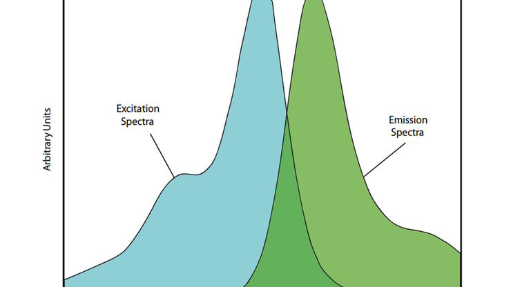
荧光染料
荧光显微镜的基本原理是借助荧光染料对细胞成分进行高度特异性的可视化观察。这可能是一种与兴趣蛋白质遗传相关的荧光蛋白,如绿色荧光蛋白(GFP)等。如果克隆无法实现,例如在组织学样本上无法实现,则需要使用另一种技术如免疫荧光染色来对兴趣蛋白质进行可视化观察。为此,人们使用抗体来连接不同的荧光染料并将其直接或间接地结合到适当的靶点上。此外,借助荧光染料,荧光显微镜的应用就不再仅局限于蛋白质观察,还能对核…
Loading...
Explore Innovative Techniques to Separate Fluorophores with Overlapping Spectra
In this article we explore several strategies you can take to improve the separation of fluorophores and increase the number of fluorescent probes you can distinguish in your sample.
Loading...
荧光和量子点的基本原理和发展历史
在您的科研生涯的某个时候,都有可能会用到荧光显微镜。这种无处不在的技术改变了显微镜学家对研究对象进行成像、标记和追踪的方式,不论是整个生物体,还是单个蛋白质等等。
通过本文,我们将探讨什么是“荧光”,包括其定义背后的历史和基础物理原理,绿色荧光蛋白(GFP)的发现和应用,并展望量子点等荧光探针不断扩大的应用领域。
Loading...
![[Translate to chinese:] Left: Tissue cells marked with an immunolabel (FITC) illuminated with wide-band UV excitation. Note the tissue structure with blue autofluorescence. Right: Same tissue and same immunostaining with FITC label illuminated with epi-illumination using narrow-band blue (490 nm) light. Note the increased image contrast (Ploem, 1967)](/fileadmin/_processed_/c/2/csm_Ploem_Figure_5_Autofluorescence_a_b_3ad909dc27.png "[Translate to chinese:] Left: Tissue cells marked with an immunolabel (FITC) illuminated with wide-band UV excitation. Note the tissue structure with blue autofluorescence. Right: Same tissue and same immunostaining with FITC label illuminated with epi-il")
Milestones in Incident Light Fluorescence Microscopy
Since the middle of the last century, fluorescence microscopy developed into a bio scientific tool with one of the biggest impacts on our understanding of life. Watching cells and proteins with the…
Loading...

Chronic Inflammation Under the Microscope
In the course of chronic inflammation certain body areas are recurrently inflamed. This goes along with many human diseases. With the help of widefield light microscopy, the underlying processes can…
Loading...
Handbook of Optical Filters for Fluorescence Microscopy
Fluorescence microscopy and other light-based applications require optical filters that have demanding spectral and physical characteristics. Often, these characteristics are application-specific and…