相同原理,两套系统
激光显微切割通常用于基因组学(DNA)、转录物组学(mRNA、miRNA)、蛋白质组学、代谢物组学,甚至下一代测序(NGS)。神经学、癌症研究、植物分析、法医学或气候研究人员均借助这种显微切割技术进行学科研究。此外,激光显微切割也是活细胞培养 (LCC) 的一款理想工具,可用于克隆、再培养、操作或下游分析。
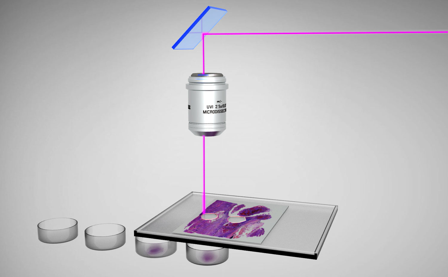
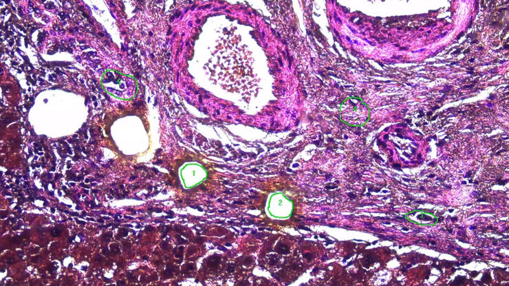
与传统激光显微切割系统不同,徕卡激光显微切割系统无需移动样品,而是通过移动激光、重力收集,大限度地避免样品污染,为您提供可即时分析的理想切割组织样品。
工作利器
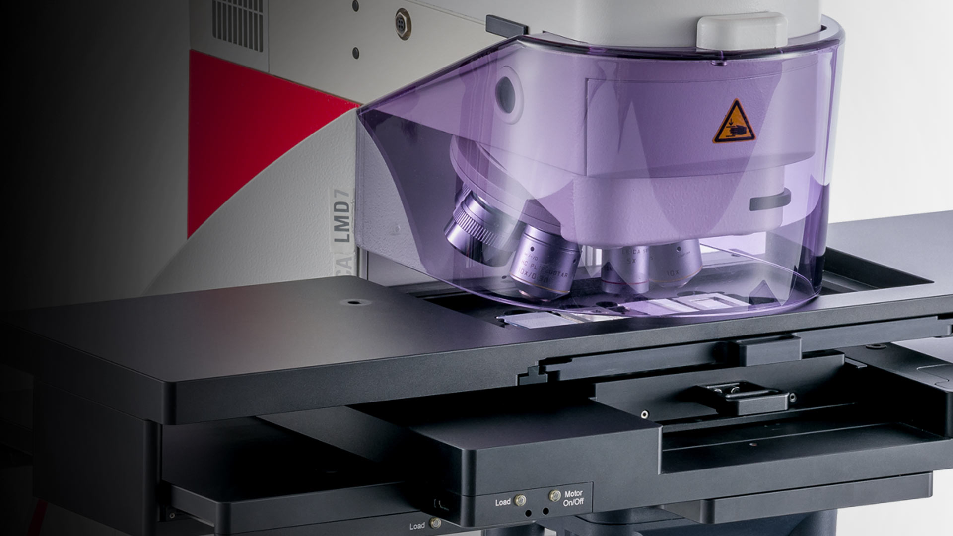
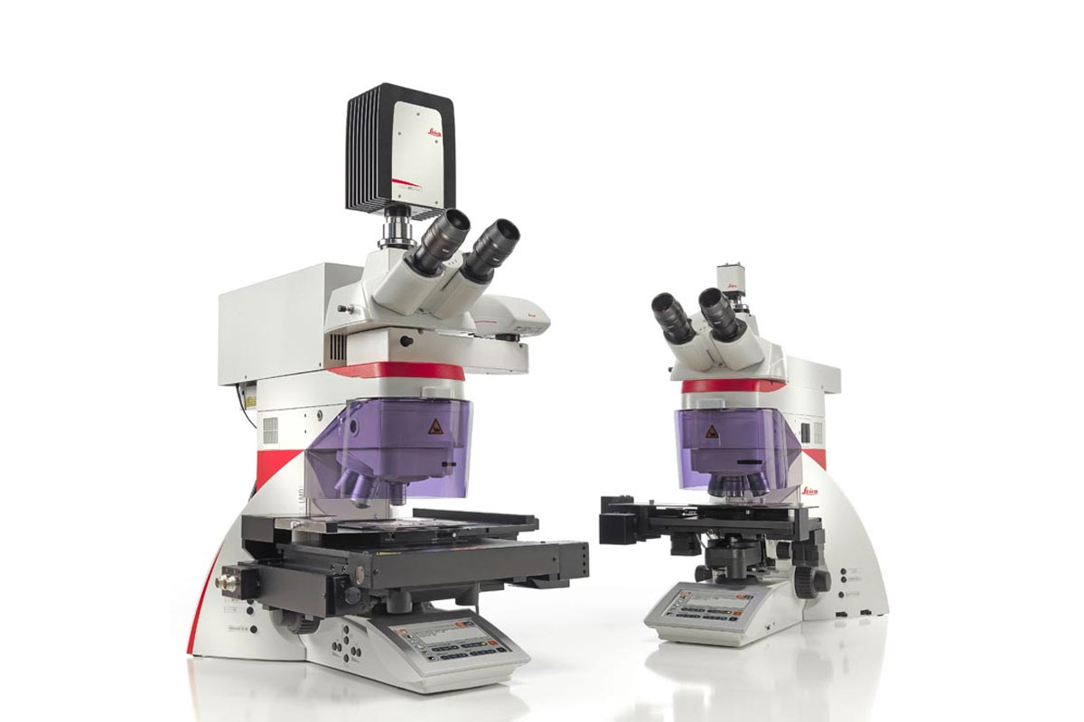
Leica LMD6 和 Leica LMD7 ,两款产品的区别主要在于激光。Leica LMD6 是解剖大脑、肝脏或肾脏等软组织标准应用的理想工具。而Leica LMD7 可以理想地切割任何类型、大小或形状的组织。与较小系统相比,Leica LMD7提供了更大的灵活性、更高的激光功率和更多的激光控件。
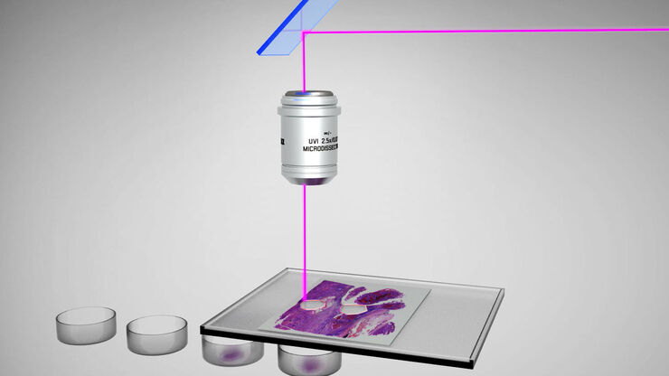
我们移动的是激光,而不是样品
当你尝试通过移动纸张而不是移动笔在一张纸上写下您的名字时,是否很难做到?是的,显然移动笔比移动纸张更快更容易,而笔就相当于我们的激光。因此,我们在激光捕获显微切割中选择移动激光,而不是移动样品。
目前,只有徕卡显微系统采用高精确度的光学部件并借助棱镜沿着组织上所需的切割线对激光束进行操纵激光束。这意味着徕卡激光显微切割可垂直于组织实施切割,从而获得切割精确、无污染的分离体。
精确无误 始终如一
- 以较高的精度和速度实施切割
- 使用“移动切割”技术,进行直接、实时地切割
- 能够获得理想视野,影像录制便利
根据您的样本调整激光
LMD7可以根据样本的需求调节能量、孔径、速度、逆流和脉冲频率的设置。例如,较厚的植物切片需要高性能,而剪切脆弱的神经元则需要较少的能量。切割薄切片时通常会比厚切片更快。
先进的切割模式可提高样本切割方法的精度。例如,您可以先绘制然后切割(绘制 + 切割),也可以通过鼠标点击或使用 PEN 屏幕进行实时切割(移动 + 切割)。绘制 + 扫描模式用于在玻璃上消融,而激光螺旋可以连续切割厚样本。使用最终脉冲模式,您可以让符合严格要求的切片掉入收集容器中。


重力收集,实现清洁无污染
下游分析需要具备无污染的分离体。因此,徕卡激光捕获显微切割系统借助重力对切除组织进行收集。其基于激光引导的独特显微切割技术保留了分离体的完整性,使其保持无接触、无污染的状态。
三步获得无污染样品:
- 选择目标区域
- 沿着要切除的区域移动激光
- 100%无污染的切除组织落入培养皿中,供进一步分析使用
真正资产:重力始终有效。
直观易用的软件使激光显微切割更方便
徕卡激光显微系统软件易于使用且功能强大,便于选择、切割和可视化切除组织,为您提供直观的切割结果。
- 可以概览样品,进行更好的定位
- 使用鼠标或触摸屏引导激光束
- 控制激光和显微镜
- 录制延时影像
- 诸如数据库、自动细胞识别 (AVC、模式识别) 等附加软件包及更多特色功能随时为您提供服务。联系徕卡销售代表 了解我们提供的全面服务!
- 终极目标:省时省力

自动化选项
ADM(自动检测模式)是帮助收集大量类似切片的软件模块。您只需标注出您感兴趣的区域,该软件就会将其用作其他区域的模板。例如,蛋白质组学分析需要采集大量样本,ADM为处理 100'-1000's 切片的实验室提供巨大的优势。
每次切片之前可以使用自动对焦,确保分布在载玻片上的数百个感兴趣区域的焦点。此外,您还可以自动检查和记录所使用的所有收集设备。
系统化方法:栅格工具将视野划分为给定数量的区域。使用此功能,您可以系统化地解剖样本,将切片收集到各种收集设备中,例如96孔板。


人工智能界面
Aivia是我们基于人工智能的图像可视化、分析和解读平台。通过人工智能增强工具(如像素分类器),Aivia可以自动定义要进行激光显微切割(LMD)的感兴趣区域(ROI)。Aivia可以检测到ROI,将其直接导入LMD软件中进行显微切割。
除了Aivia之外,还可以使用其他外部软件自动检测ROI。LMD软件只需要一个包含ROI信息的XML文件。
- Mund et al., Nature Biotechnology, 2022 https://doi.org/10.1038/s41587-022-01302-5
- Mitchell et al., JoVE, 2022 https://doi.org/10.3791/64171

高处理能力
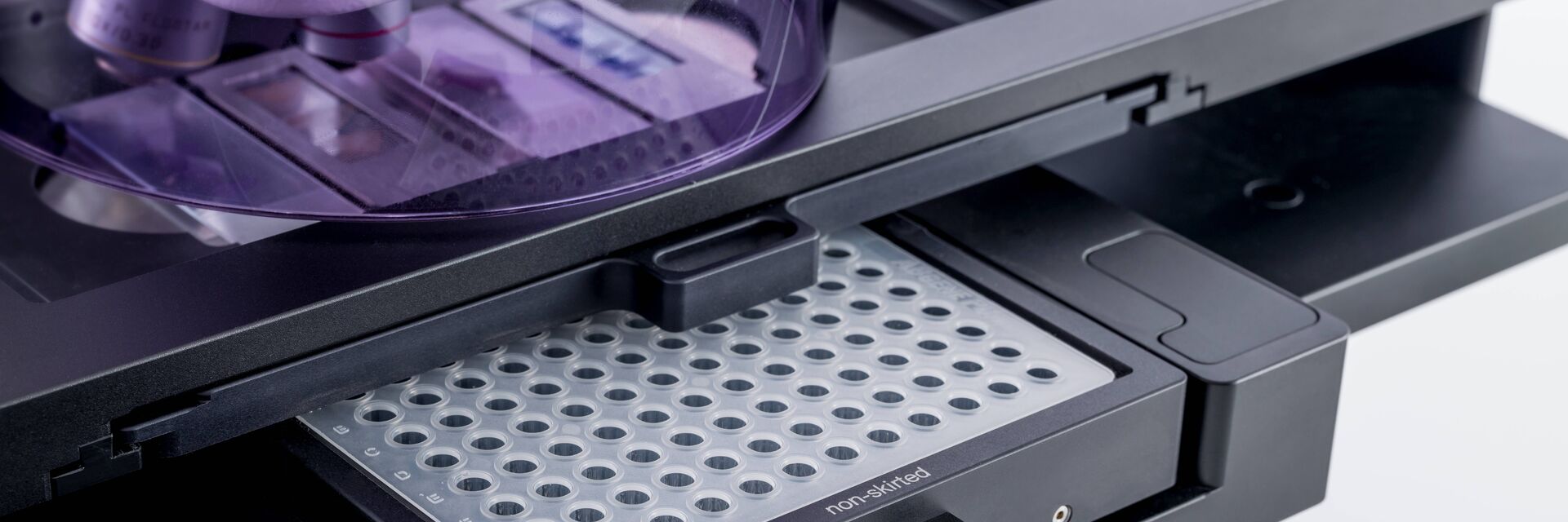
需要提高处理量?LMD系统可以配置不同的载物台。其中,LMT350扫描载物台可提供最快、最安静、最精确的导航。
LMT350中能够放置0.2毫升带帽管、8联和12联排管以及标准96孔板与384孔板*。这使您能够进行高通量实验。
因为您不需要任何特殊材料,只需要使用常规的微量滴定板,这些孔板可以直接装在标准PCR机器上。
*最多可处理352孔
均匀照明
光线在界定切割区域时至关重要。Leica LMD6 和 Leica LMD7 激光显微切割系统可采用传统卤素灯或 LED 照明。
LED 照明为您带来的好处:
- 在充足的照明下,您可以看到样品的自然颜色 - 因为 LED 照明可均匀照亮样品,且具有恒定的色温
- LED 照明可节省 90% 的能源,并具有长达 25,000 小时的使用寿命 –因更换灯泡而导致的仪器停机早已成为过去式
- 如果用于透射光的卤素照明仍然是您的优选,我们的这两种系统也均可配备。我们可为其提供内部恒定色温控制 (CCIC),以避免由于采用传统照明技术而导致的任何图像变化,即使将系统用于与激光显微切割无关的应用也没有问题
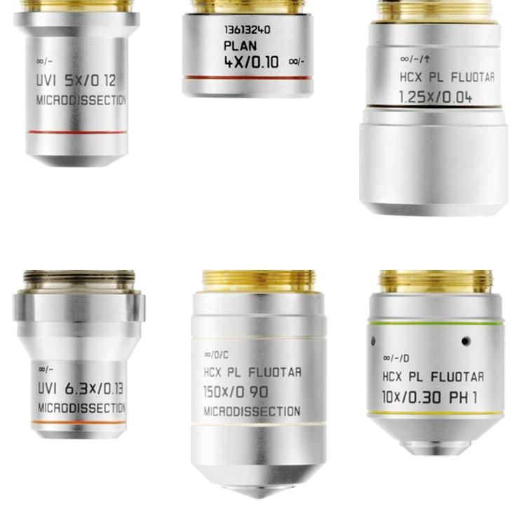
性能优异的物镜
从19 世纪初,光学部件的开发和制造就已经是我们核心竞争力的一个重要部分。请信赖徕卡SmartCut系列激光显微切割专用物镜的优良性能,它帮您实现优异切割效果!
- 选择范围:10 种干式物镜, 从 5x 到 150x
- 出色的成像性能,需要时,还可采用独特的 150x SmartCut 物镜观察到高放大倍率、高分辨率下的样品细节
- 使用低放大倍率物镜可获得更大的视场,以完好无损地切割大块样品
- 物镜激光透光率可达 350 nm,可完成对组织、骨骼、牙齿、大脑、植物、染色体和活细胞的激光显微切割
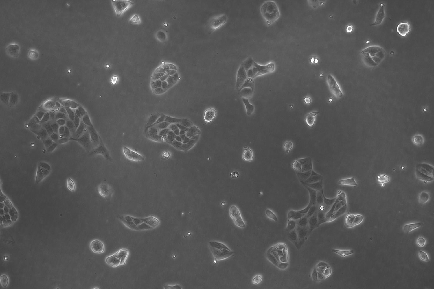
便于活细胞切片
徕卡激光显微切割系统,采用立式光学成像系统,为您提供高分辨率视野,确保对活体组织、细胞乃至亚细胞结构的精确区分与切割。
- 您可以切割培养菌中的活细胞,以重新培养、克隆或分析单个细胞、菌落或细胞群
- 您甚至可以将气候室连接到激光显微切割系统
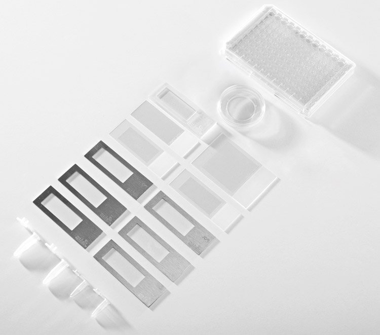
- 您可使用 PEN 薄膜或多皿 ibidi 载玻片在培养皿中培植细胞
- 您可将活细胞培养菌的切除组织收集到培养皿 (带或不带 PEN 薄膜、ibidi 载玻片、或 8 条纹管均可) 中重新培养,或者收集到 PCR 管帽等收集设备中进行分析
二合一系统:LMD 系统与THUNDER成像系统相结合
LMD6 和 LMD7 系统可与 THUNDER 组合使用。THUNDER Imager 3D Tissue 和LMD系统具有同一个底座,因此这样的组合具有以下优点:
- 用一个系统完成不同的任务,节省实验室空间
- 用LMD不受任何限制地进行激光显微切割
- 用LAS X进行出色的THUNDER荧光成像,包括3D查看器中的多色3D(Z轴层扫)显示。


