
Loading...
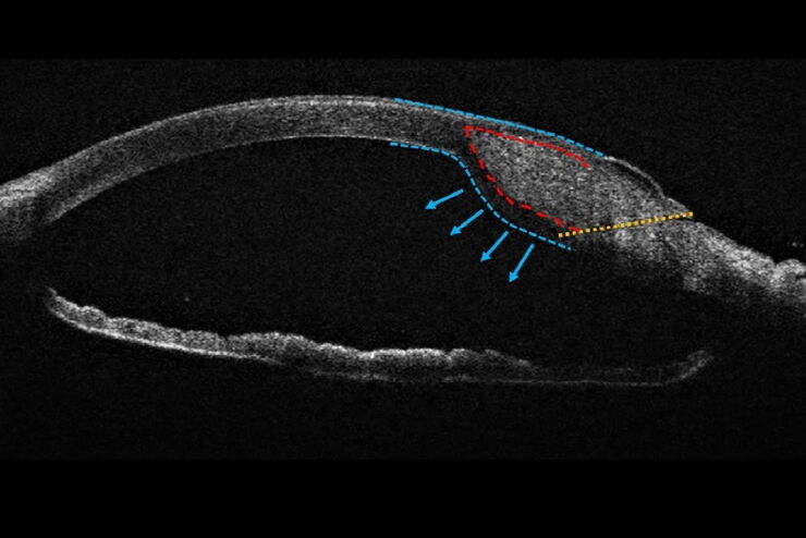
Towards Advanced Use of Intraoperative OCT in Cataract Surgery
In this White Paper, Dr. Rachid Tahiri shares his personal experience with the Leica EnFocus intraoperative OCT, the valuable features supporting smooth surgery and how it allows him to minimize…
Loading...
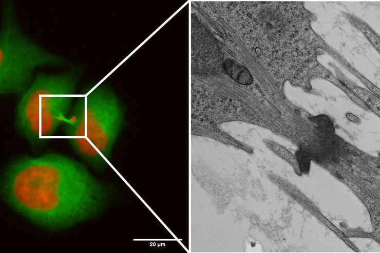
Capture life as it happens
With the Leica Nano Workflow, searching for the needle in the haystack is a thing of the past. Take advantage of correlative light and electron microscopy to identify directly the right cell at the…
Loading...
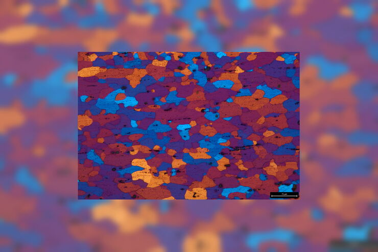
包括成分分析在内的微观结构表征
Leica Microsystems的多功能正置复合显微镜DM6M配有激光诱导击穿光谱模块,不仅可以分析金相抛光样品,进行晶粒度分析和钢铁夹杂物评级,而且在同一系统上,同时获得关于小到20微米的化学成分的定性组成信息,而无需任何额外的样品制备并且无需将样品转移到电子显微镜。加入我们,看看这个系统的运作。
Loading...

Life Beyond the Pixels: Deep Learning Methods for Single Cell Analysis
Our guest speaker Prof Dr Peter Horvath presents his work on single cell-based large-scale microscopy experiments. This novel targeting approach includes the use of machine learning models and…
Loading...
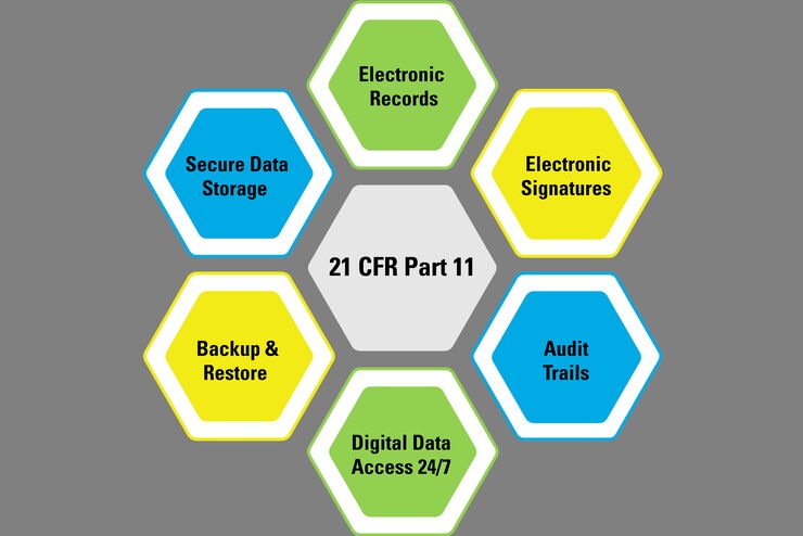
美国联邦法规第21章第11款和其他相关法规简介
本文概述了在美国(联邦法规第21章第11款)、欧盟(GMP附录11)和中国(NMPA)所用电子记录(数据输入、存储、签名和审批)的法规和指南,这些法规和指南会对医疗器械质量控制的数字化增强检测解决方案产生影响。与纸质记录方法相比,使用显微镜进行数字化增强检测具有更一致和更高效的检测优势。但是,与纸质记录和签名的规定相比,电子记录和签名的规定有明显不同的建议和要求。电子记录的创建、验证、存储和备份应…
Loading...

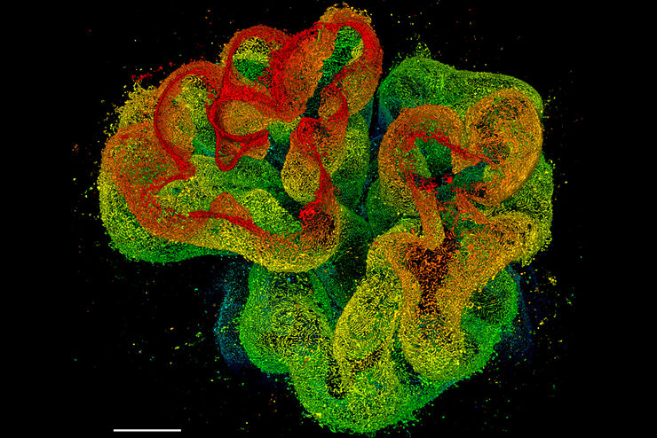
Physiology Image Gallery
Physiology is about the processes and functions within a living organism. Research in physiology focuses on the activities and functions of an organism’s organs, tissues, or cells, including the…
Loading...
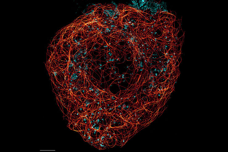
超分辨率显微镜图片库
由于光的衍射极限,传统共聚焦显微镜无法分辨约240纳米以下的结构。当需要提高分辨率以研究衍射极限尺度以下的结构和分子事件时,会使用超分辨率显微镜技术,如STED、PALM或STORM,或某些解卷积处理方法。
Loading...
组织图片库
对动物和人体组织进行视觉分析对于了解癌症或神经变性等复杂疾病至关重要。从基本的免疫组化到体内成像,共聚焦显微镜和先进的模式可以让人们了解细胞、生物分子及其在环境中的相互作用。
Loading...
![[Translate to chinese:] Nematostella](/fileadmin/_processed_/d/c/csm_Nematostella-LiveImaging-Stellaris_1d96dd4af5.jpg "[Translate to chinese:] Nematostella")
活细胞成像图库
活细胞显微镜技术是更好地了解细胞和分子功能的基础。如今,宽场显微镜是用于长时间观察细胞动态和发育的最常用技术。共聚焦显微镜也是一种重要工具,可生成三维结构图像,并以高空间和时间分辨率研究高度动态的细胞过程,同时使标本保持接近原生状态。