
Loading...

What are the Challenges in Neuroscience Microscopy?
eBook outlining the visualization of the nervous system using different types of microscopy techniques and methods to address questions in neuroscience.
Loading...
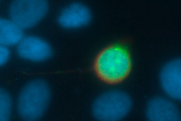
![[Translate to chinese:] Raw widefield and THUNDER image of calcium transients in Drosophila embryos. Courtesy A. Carreira-Rosario, Clandinin laboratory, California, USA.](/fileadmin/_processed_/7/6/csm_Calcium_transients_in_Drosophila_embryos_teaser_fcd31bd78f.jpg "[Translate to chinese:] Raw widefield and THUNDER image of calcium transients in Drosophila embryos. Courtesy A. Carreira-Rosario, Clandinin laboratory, California, USA.")
Central Nervous System (CNS) Development and Activity in Organisms
This article shows how studying central nervous system (CNS) development in Drosophila-melanogaster embryos expressing a GCaMP calcium indicator in the neurons can be improved with a THUNDER Imager.
Loading...
Going Beyond Deconvolution
Widefield fluorescence microscopy is often used to visualize structures in life science specimens and obtain useful information. With the use of fluorescent proteins or dyes, discrete specimen…
Loading...

快速、高灵敏度成像和人工智能辅助分析
The specificity of fluorescence microscopy allows researchers to accurately observe and analyze biological processes and structures quickly and easily, even when using thick or large samples. However,…
Loading...

为活细胞成像创造新选择
对厚实的活体样本进行成像时,主要挑战之一是获得图像质量与组织完整性之间的平衡。长时间的图像采集期间,弱信号光会导致低信号水平,导致图像对比度低以及分割和分析困难。需要通过高剂量成像或高时间分辨率成像技术加强信号强度时,这一问题更加突出。一个常见问题是:我如果快速成像、一次完成,会不会造成样本过度漂白或者细胞死亡?
Loading...

精确分析宽视野荧光图像
利用荧光显微镜的特异性,即便是使用厚样品和大尺寸样品,研究人员也能够快速轻松地准确观察和分析生物学过程和结构。然而,离焦荧光会提高背景荧光,降低对比度,影响图像的精确分割。THUNDER 与Aivia 的组合可以有效解决这一问题。前者可以消除图像模糊,后者会使用人工智能技术自动分析宽视野图像,提高操作速度和精确性。下面,我们来详细了解下这一协作方法。
Loading...

High-resolution 3D Imaging to Investigate Tissue Ageing
Award-winning researcher Dr. Anjali Kusumbe demonstrates age-related changes in vascular microenvironments through single-cell resolution 3D imaging of young and aged organs.
Loading...
Optimizing THUNDER Platform for High-Content Slide Scanning
With rising demand for full-tissue imaging and the need for FL signal quantitation in diverse biological specimens, the limits on HC imaging technology are tested, while user trainability and…
Loading...

Physiology Image Gallery
Physiology is about the processes and functions within a living organism. Research in physiology focuses on the activities and functions of an organism’s organs, tissues, or cells, including the…