
Loading...

A Meta-cancer Analysis of the Tumor Spatial Microenvironment
Learn how clustering analysis of Cell DIVE datasets in Aivia can be used to understand tissue-specific and pan-cancer mechanisms of cancer progression
Loading...
 tissue.")
Mapping the Landscape of Colorectal Adenocarcinoma with Imaging and AI
Discover deep insights in colon adenocarcinoma and other immuno-oncology realms through the potent combination of Cell DIVE's multiplexed imaging and Aivia AI-based image analysis
Loading...

Spatial Architecture of Tumor and Immune Cells in Tumor Tissues
Dig deep into the spatial biology of cancer progression and mouse immune-oncology in this poster, and learn how tumor metabolism can effect immune cell function.
Loading...

IBEX, Cell DIVE, and RNA-Seq: A Multi-omics Approach to Follicular Lymphoma
In a recent study by Radtke et al., a multi-omics spatial biology approach helps shed light on early relapsing lymphoma patients
Loading...

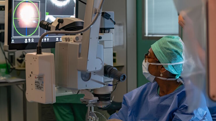
Dislocated Cataract Angle Closure Aided by Intraoperative OCT
Learn how a dislocated cataract was treated with angle closure assisted by intraoperative OCT to achieve long-term good results without future lens dislocation.
Loading...
![[Translate to chinese:] Adult human Alzheimer’s brain demonstrating a panel of 15 markers.](/fileadmin/_processed_/4/9/csm_Adult_human_Alzheimers_brain_68bacde06e.jpg "[Translate to chinese:] Adult human Alzheimer’s brain demonstrating a panel of 15 markers.")
大脑的形状:阿尔茨海默病的空间生物学
阿尔茨海默病(AD)是一种神经退行性疾病,也是导致晚年认知障碍的常见原因。阿尔茨海默病的特征是出现含β-淀粉样蛋白的斑块和含磷酸化 tau 的神经纤维缠结。目前尚缺乏治疗和预防AD的有效疗法。我们将Cell DIVE与Cell Signaling Technology的抗体结合使用,检查了AD的突触过程并从空间上确定了神经胶质细胞和神经元等细胞,证明了超多标免疫荧光成像技术可用于探测AD大脑。
Loading...

术中OCT引导的青光眼支架修复手术
青光眼是导致全球不可逆失明的主要原因之一。小梁网切除术和导管分流引流术等历史悠久的手术技术会带来巨大的短期风险和潜在并发症。近年来,随着微创青光眼手术(MIGS)的出现,手术方法有了长足的发展,其特点是对组织的破坏最小、内路粘小管植入、手术时间短、器械简单、术后恢复快。
Loading...
![[Translate to chinese:] Co-detection of 10 extracellular matrix proteins and 3 topographical tissue landmarks by multiplex immunostaining within a single high-grade fibrous hotspot from a human hepatocellular carcinoma](/fileadmin/_processed_/0/8/csm_Single_high-grade_fibrous_hotspot_from_a_human_hepatocellular_carcinoma_e5541282bd.jpg "[Translate to chinese:] Co-detection of 10 extracellular matrix proteins and 3 topographical tissue landmarks by multiplex immunostaining within a single high-grade fibrous hotspot from a human hepatocellular carcinoma")
肝细胞癌中癌症干细胞位点的原位鉴定
在这里,我们探索了一种突破性的多重免疫检测方法,通过多重成像对细胞外基质(ECM)特征进行原位定位,从而识别肝细胞癌(HCC)内的癌症干细胞龛。