
Loading...

High-resolution 3D Imaging to Investigate Tissue Ageing
Award-winning researcher Dr. Anjali Kusumbe demonstrates age-related changes in vascular microenvironments through single-cell resolution 3D imaging of young and aged organs.
Loading...
透明介质对组织透明度和收缩的影响
本研究通过比较新鲜解剖的双翅目昆虫脑与其透明化处理后的等效物,全面评估了不同透明化介质对组织透明度和收缩率的影响。组织透明化处理结合光片显微技术,已成为全器官三维成像和定量的强大工具。由于组织透明化处理有助于在亚细胞水平上对完整组织进行光学成像,它有潜力揭示如大脑等复杂器官以前未见的细节。为了对厚样本进行高分辨率成像,透明化介质需要与高数值孔径(NA)物镜使用的浸没液具有匹配的高折射率。这一点对于…
Loading...
![[Translate to chinese:] Elucidate cancer development on sub-cellular level by in-vivo like tumor spheroid models.](/fileadmin/_processed_/f/5/csm_3d-biology-workflow-DLS_3e7765c385.jpg "[Translate to chinese:] Elucidate cancer development on sub-cellular level by in-vivo like tumor spheroid models.")
利用光片显微镜改进三维细胞生物学工作流程
了解癌症发生过程中的亚细胞机制对于癌症治疗至关重要。常见的细胞模型涉及作为单层生长的癌细胞。然而,这种方法忽视了肿瘤细胞与其周围微环境之间的三维相互作用。为了贴近自然环境理解恶性肿瘤的发展和进程,对癌症微环境的详细表征至关重要。
![[Translate to chinese:] Array tomography image of T-cells in mouse lymph nodes.](/fileadmin/_processed_/b/9/csm_T-cells_in_mouse_lymph_nodes_array_tomography_d9e5d6f4da.jpg "[Translate to chinese:] Array tomography image of T-cells in mouse lymph nodes.")
Loading...
BABB Clearing and Imaging for High Resolution Confocal Microscopy
Multipohoton microscopy experiment using Leica TCS SP8 MP and Leica 20x/0.95 NA BABB immersion objective.
Understanding kidney microanatomy is key to detecting and identifying early events in kidney…
Loading...

Confocal and Light Sheet Imaging with STELLARIS DLS
Optical imaging instrumentation can magnify tiny objects, zoom in on distant stars and reveal details that are invisible to the naked eye. But it notoriously suffers from an annoying problem: the…
Loading...
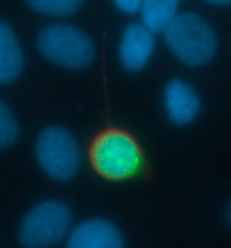
Image Processing for Widefield Microscopy
Fluorescence microscopy is a modern and steadily evolving tool to bring light to current cell biological questions. With the help of fluorescent proteins or dyes it is possible to make discrete…