
Loading...
![[Translate to chinese:] Co-detection of 10 extracellular matrix proteins and 3 topographical tissue landmarks by multiplex immunostaining within a single high-grade fibrous hotspot from a human hepatocellular carcinoma](/fileadmin/_processed_/0/8/csm_Single_high-grade_fibrous_hotspot_from_a_human_hepatocellular_carcinoma_e5541282bd.jpg "[Translate to chinese:] Co-detection of 10 extracellular matrix proteins and 3 topographical tissue landmarks by multiplex immunostaining within a single high-grade fibrous hotspot from a human hepatocellular carcinoma")
肝细胞癌中癌症干细胞位点的原位鉴定
在这里,我们探索了一种突破性的多重免疫检测方法,通过多重成像对细胞外基质(ECM)特征进行原位定位,从而识别肝细胞癌(HCC)内的癌症干细胞龛。

Loading...

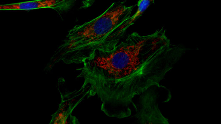
Epi-Illumination Fluorescence and Reflection-Contrast Microscopy
This article discusses the development of epi-illumination and reflection contrast for fluorescence microscopy concerning life-science applications. Much was done by the Ploem research group…
Loading...

Discover how Multiplexed Bioimaging can Advance Cancer Research
Explore multiplexing with up to 60 biomarkers, enabling advanced tumor imaging approaches to gather precise, spatially-resolved single-cell data that helps enhance cancer research and clinical…
![[Translate to chinese:] Prof. Nikolaos Bechrakis uses the Proveo 8 ceiling mounted microscope with EnFocus intraoperative OCT. Images provided by Prof. Nikolaos Bechrakis.](/fileadmin/_processed_/7/3/csm_Prof_Bechrakis_uses_Proveo_8_ceiling_mounted_microscope_with_EnFocus_intraoperative_OCT_dad0be1120.jpg "[Translate to chinese:] Prof. Nikolaos Bechrakis uses the Proveo 8 ceiling mounted microscope with EnFocus intraoperative OCT. Images provided by Prof. Nikolaos Bechrakis.")
Loading...
![[Translate to chinese:] Material sample with a large height, size, and weight being observed with an inverted microscope.](/fileadmin/_processed_/f/4/csm_Inverted_microscope_with_large_sample_crop_04c3257c81.jpg "[Translate to chinese:] Material sample with a large height, size, and weight being observed with an inverted microscope.")
工业应用中倒置显微镜相较于正置显微镜的五大优势
使用倒置显微镜时,您需要从下方观察样本,因为倒置显微镜的光学元件位于样本下方,而使用正置显微镜时,您需要从上方观察样本。一直以来,倒置显微镜主要用于生命科学研究,因为重力将样本沉入含有水性溶液的托座底部,从上方则无法观察到太多内容。但近段时间以来,倒置显微镜在工业应用中也变得越来越流行。我们现在一起来了解倒置显微镜在工业应用中的优势。
Loading...

共聚焦多色成像在癌症研究和免疫学中的潜力
在本次网络研讨会上,来自莫纳什制药科学研究所的CameronNowell和他的同事将分享他们在多重成像方面的经验,以及他们通过巧妙的共聚焦成像采集和利用FLIM等其他多重成像模式所取得的成果。
![[Translate to chinese:] Keratoplasty of pathologic cornea](/fileadmin/_processed_/6/d/csm_Pathologic_cornea_cb99c6735b.jpg "[Translate to chinese:] Keratoplasty of pathologic cornea")