
Loading...
![[Translate to chinese:] Augmented Reality fluorescence supports each step of neurovascular surgery procedures. Image courtesy of Dr. Christof Renner.](/fileadmin/_processed_/4/f/csm_Augmented_Reality_fluorescence_supports_neurovascular_surgery_procedures_af3ba59a73.jpg "[Translate to chinese:] Augmented Reality fluorescence supports each step of neurovascular surgery procedures. Image courtesy of Dr. Christof Renner.")
AR荧光在神经血管手术中的应用
术中血管造影在神经血管手术中发挥着至关重要的作用。在徕卡2021神经可视化峰会期间,Christof Renner博士在独家网络研讨会上展示了精选临床病例,并分享了使用GLOW800增强现实荧光技术的经验。
Loading...
![[Translate to chinese:] C. elegans adult hermaphrodite gonades acquired using THUNDER Imager. Staining: blue - DAPI (nucleus), green - SP56 (sperm), red - RME-2 (oocyte), magenta - PGL-1 (RNA + protein granules). Image courtesy of Prof. Dr. Christian Eckmann, Martin Luther University, Halle, Germany.](/fileadmin/_processed_/7/c/csm_C_elegans_adult_hermaphrodite_gonades_6194ed03fc.jpg "[Translate to chinese:] C. elegans adult hermaphrodite gonades acquired using THUNDER Imager. Image courtesy of Prof. Dr. Christian Eckmann, Martin Luther University, Halle, Germany.")
生命科学研究: 哪种显微镜相机适合您?
相机是显微镜系统的重要组成部分,对系统的性能有重大影响。在选择相机时,重要的是不仅要看技术规格,还要考虑您的样品、技术、对比方法以及您希望获得的数据类型。
Loading...
![[Translate to chinese:] Pancreatic Ductal Adenocarcinoma with 11 Apoptosis biomarkers shown – BAK, BAX, BCL2, BCLXL, Caspase9, CIAP1, NaKATPase, PCK26, SMAC, Vimentin, and XIAP.](/fileadmin/academy/2023/Gated_content/Pancreatic_Ductal_Adenocarcinoma_11_Apoptosis_Markers_ROI5.jpg "[Translate to chinese:] Pancreatic Ductal Adenocarcinoma with 11 Apoptosis biomarkers shown – BAK, BAX, BCL2, BCLXL, Caspase9, CIAP1, NaKATPase, PCK26, SMAC, Vimentin, and XIAP.")
与卢克-加蒙(Luke Gammon)一起多重成像:推进您的空间生物学研究
多重成像是一种功能强大的技术,可让研究人员同时观察单个样本中的多个目标。这对于研究复杂的生物系统尤为重要,可以帮助研究人员更好地了解不同分子和途径之间是如何相互作用的。
Loading...

Spatial Biology: Learning the Landscape
Spatial Biology: Understanding the organization and interaction of molecules, cells, and tissues in their native spatial context
Loading...

考虑采购体视显微镜时的关键因素
体视显微镜通常是实验室或生产现场“主力”。用户需要花费数小时通过目镜来检查、观察、记录或解剖样本。仔细评估哪些相关应用需要用到体视显微镜,是确保长期满意使用的关键所在。决策者们需要确保自己能够完全依照自己的需求来定制仪器。为帮助用户能更好的选择适合自己的体视镜,本文介绍了几个主要考虑的因素。
Loading...
![[Translate to chinese:] Brain organoid section (DAPI) acquired using THUNDER Imager Live Cell. Image courtesy of Janina Kaspar and Irene Santisteban, Schäfer Lab, TUM.](/fileadmin/_processed_/2/7/csm_Tilescan_of_brain_organoid_section_46d510ba4e.jpg "[Translate to chinese:] Brain organoid section (DAPI) acquired using THUNDER Imager Live Cell. Image courtesy of Janina Kaspar and Irene Santisteban, Schäfer Lab, TUM.")
研究大脑健康的成像类器官模型
小胶质细胞是特化的脑驻留免疫细胞,在大脑发育、平衡和疾病中发挥着至关重要的作用。然而,到目前为止,模拟人脑环境与小胶质细胞之间相互作用的能力还非常有限。
Loading...
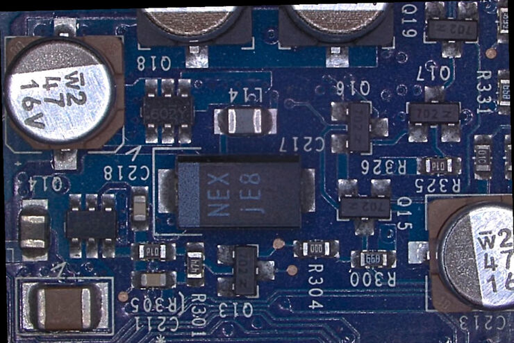
利用数码显微镜快速、可靠地对印刷电路板(PCB)及其总成(PCBA)进行显微观察
本文阐释了徕卡数码显微镜DVM6的性能优势,例如简单直观的操作系统、快速简单的放大倍率切换方式,并且可以通过编码准确调取参数。
![[Translate to chinese:] In vivo imaging of a mouse pial and cortical vasculature through a glass window (ROSAmT/mG::Pdgfb-CreERT2 mouse meningeal and cortical visualization following tamoxifen induction and craniotomy). Courtesy: Thomas Mathivet, PhD](/fileadmin/_processed_/6/3/csm_Mouse_pial_and_cortical_vasculature_f409d941d0.jpg "[Translate to chinese:] In vivo imaging of a mouse pial and cortical vasculature through a glass window (ROSAmT/mG::Pdgfb-CreERT2 mouse meningeal and cortical visualization following tamoxifen induction and craniotomy). Courtesy: Thomas Mathivet, PhD")
Loading...

Ophthalmology Case Study: Corneal Transplantation
Learn about the use of intraoperative Optical Coherence Tomography in Corneal Transplantation and how it helps achieve correct positioning of donor tissue.