
Loading...

Find Relevant Specimen Details from Overviews
Switch from searching image by image to seeing the full overview of samples quickly and identifying the important specimen details instantly with confocal microscopy. Use that knowledge to set up…
Loading...

精确分析宽视野荧光图像
利用荧光显微镜的特异性,即便是使用厚样品和大尺寸样品,研究人员也能够快速轻松地准确观察和分析生物学过程和结构。然而,离焦荧光会提高背景荧光,降低对比度,影响图像的精确分割。THUNDER 与Aivia 的组合可以有效解决这一问题。前者可以消除图像模糊,后者会使用人工智能技术自动分析宽视野图像,提高操作速度和精确性。下面,我们来详细了解下这一协作方法。
Loading...
Save Time and Effort with AI-assisted Fluorescence Image Analysis
The powerful synergy of THUNDER and Aivia analyze fluorescence images with greater accuracy, even when using low light excitation.
Loading...

High-resolution 3D Imaging to Investigate Tissue Ageing
Award-winning researcher Dr. Anjali Kusumbe demonstrates age-related changes in vascular microenvironments through single-cell resolution 3D imaging of young and aged organs.
Loading...
![[Translate to chinese:] HeLa Kyoto cells (HKF1, H2B-mCherry, alpha Tubulin, mEGFP). Left image: Maximum projection of a z-stack prior to ICC and LVCC. Right image: Maximum projection of a mosaic z-stack after ICC and LVCC.](/fileadmin/_processed_/6/a/csm_How_to_improve_live_cell_imaging_with_Leica_Nano_Workflow_teaser_04b9da73e4.jpg "[Translate to chinese:] HeLa Kyoto cells")
如何使用Coral Life(活细胞光电联用)改进活细胞成像
对于活细胞 CLEM 应用而言,光学显微镜成像是在正确的时间以正确的状态识别正确细胞的关键步骤。在本文中,徕卡专家就使用宽场系统的优势以及使用蓝宝石作为细胞培养基底时需要克服的障碍分享了他们的见解。
Loading...
Optimizing THUNDER Platform for High-Content Slide Scanning
With rising demand for full-tissue imaging and the need for FL signal quantitation in diverse biological specimens, the limits on HC imaging technology are tested, while user trainability and…
Loading...

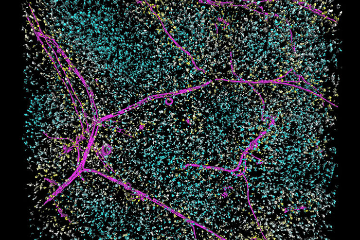
Physiology Image Gallery
Physiology is about the processes and functions within a living organism. Research in physiology focuses on the activities and functions of an organism’s organs, tissues, or cells, including the…
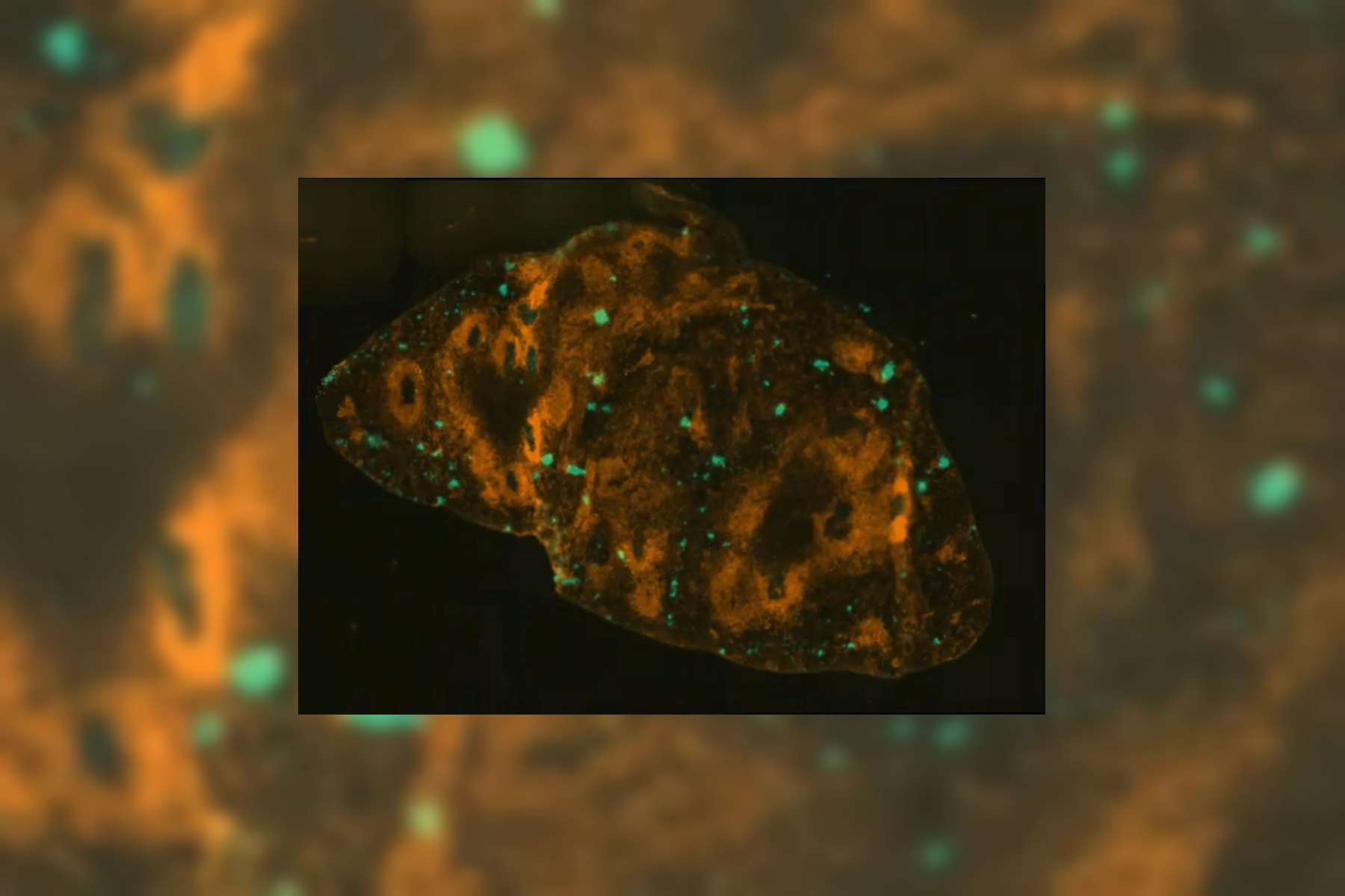
![[Translate to chinese:] Virally labeled neurons (red) and astrocytes (green) in a cortical spheroid derived from human induced pluripotent stem cells. THUNDER Model Organism Imagerwith a 2x 0.15 NA objective at 3.4x zoomwas used to produce this 425 μm Z-stack (26 positions), which is presented here as an Extended Depth of Field(EDoF)projection.](/fileadmin/_processed_/e/c/csm_THUNDER_Imager_Model-Org_Header-Gallery-Neuroscience_4652e132e6.jpg "[Translate to chinese:] Virally labeled neurons (red) and astrocytes (green) in a cortical spheroid derived from human induced pluripotent stem cells.")
Loading...

Developmental Biology Image Gallery
Developmental biology explores the development of complex organisms from the embryo to adulthood to understand in detail the origins of disease. This category of the gallery shows images about…