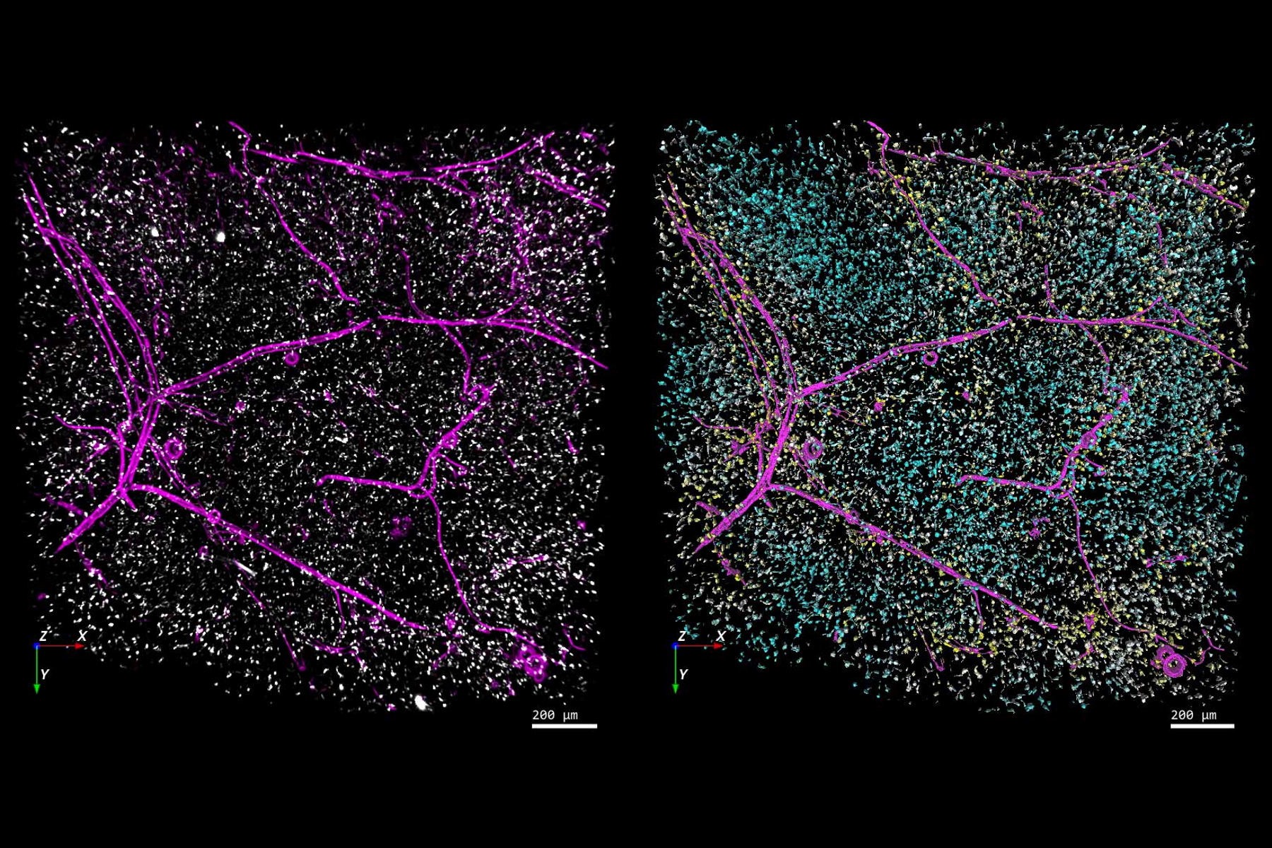
联系我们

精确分析宽视野荧光图像

如何消除离焦模糊,提高分割精确性以及在图像分析中利用人工智能技术

[Translate to chinese:] Left-hand image: The distribution of immune cells (white) and blood vessels (pink) in white adipose tissue (image captured using the THUNDER Imager 3D Cell Culture). Right-hand image: The same image after automated analysis using Aivia, with each immune cell color-coded based on its distance to the nearest blood vessel. Image courtesy of Dr. Selina Keppler, Munich, Germany. Fat_tissue_THUNDER_THUNDER-Aivia_teaser.jpg

利用荧光显微镜的特异性,即便是使用厚样品和大尺寸样品,研究人员也能够快速轻松地准确观察和分析生物学过程和结构。然而,离焦荧光会提高背景荧光,降低对比度,影响图像的精确分割。THUNDER 与Aivia 的组合可以有效解决这一问题。前者可以消除图像模糊,后者会使用人工智能技术自动分析宽视野图像,提高操作速度和精确性。下面,我们来详细了解下这一协作方法。

相关文章

  • Mouse whole-mount retina. Image courtesy of the Experimental Ophthalmology Group, University of Murcia, Spain.

    快速、高灵敏度成像和人工智能辅助分析

    The specificity of fluorescence microscopy allows researchers to accurately observe and analyze…

    Jun 01, 2022
    Read article
  • 3D reconstruction of an isolated human islet

    为活细胞成像创造新选择

    对厚实的活体样本进行成像时,主要挑战之一是获得图像质量与组织完整性之间的平衡。长时间的图像采集期间,弱信号光会导致低信号水平,导致图像对比度低以及分割和分析困难。需要通过高剂量成像或高时间分辨率成像技…

    Apr 08, 2022
    Read article
  • Left-hand image: The distribution of immune cells (white) and blood vessels (pink) in white adipose tissue (image captured using the THUNDER Imager 3D Cell Culture). Right-hand image: The same image after automated analysis using Aivia, with each immune cell color-coded based on its distance to the nearest blood vessel. Image courtesy of Dr. Selina Keppler, Munich, Germany.

    精确分析宽视野荧光图像

    利用荧光显微镜的特异性,即便是使用厚样品和大尺寸样品,研究人员也能够快速轻松地准确观察和分析生物学过程和结构。然而,离焦荧光会提高背景荧光,降低对比度,影响图像的精确分割。THUNDER 与Aivia…

    Jan 17, 2022
    Read article

相关页面

  • 显微镜成像软件

    徕卡显微系统的显微镜成像软件将显微镜、数码相机和各种配件结合在一个完整、综合的解决方案中。

    Visit related page
  • THUNDER Imaging Systems

    为了解答重要的科研问题,这些系统甚至能深入原始样品中实时呈现清晰的细节,不会产生任何离焦模糊。现如今,为3D样品进行清晰成像就像使用您最喜爱的摄像头荧光显微镜一样简单。采用 Computational…

    Visit related page
Background image
Scroll to top