
Loading...

Developmental Biology Image Gallery
Developmental biology explores the development of complex organisms from the embryo to adulthood to understand in detail the origins of disease. This category of the gallery shows images about…
Loading...
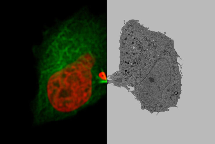
将动态活细胞数据融入超微结构
采用徕卡Nano的工作流程,可以避免过去如海底捞针似的寻找。利用光电关联显微技术,在适当的时间直接鉴别出正确的细胞,并将动态的活细胞数据融入其超微结构中。
Loading...
![[Translate to chinese:] Electroporated nerve cells (green), specific neuronal markers (magenta) and cell nuclei (white), computational cleared.](/fileadmin/_processed_/a/0/csm_Ferret_Brain_LVCC_30911a5848.jpg "[Translate to chinese:] Electroporated nerve cells (green), specific neuronal markers (magenta) and cell nuclei (white), computational cleared.")
利用 "Wow效应 "进入三维空间--以三维方式实时观察细胞
生命是瞬息万变的,对细胞来说更是如此。通常,细胞应在尽可能接近其自然环境的生理条件下进行检测。新技术为基于相机的荧光系统提供了巨大的性能,可在一次拍摄中实现全分辨率操作。本文介绍了如何利用新技术实时有效地去除焦平面以外区域不需要的图像内容。文章认为,这些新方法和数据交换正在推动科学进步。
![[Translate to chinese:] Virally labeled neurons (red) and astrocytes (green) in a cortical spheroid derived from human induced pluripotent stem cells. THUNDER Model Organism Imager with a 2x 0.15 NA objective at 3.4x zoom was used to produce this 425 µm Z-stack (26 positions), which is presented here as an Extended Depth of Field (EDoF) projection. Images courtesy of Dr. Fikri Birey from the Dr. Sergiu Pasca laboratory at Stanford University, 3165 Porter Dr., Palo Alto, CA](/fileadmin/_processed_/2/0/csm_Neural-sphere_model-org_LVC_61ecf44e40.jpg "[Translate to chinese:] Virally labeled neurons (red) and astrocytes (green) in a cortical spheroid derived from human induced pluripotent stem cells.")
Loading...
 and cytokinesis ring (Rlc1-mCherry; red).")
Studying Cell Division
Cell division is a biological process during which all cellular components must be distributed among the daughter cells. The division process requires firm coordination for success. Microscopy is…
Loading...
![[Translate to chinese:] Mouse kidney section with Alexa Fluor™ 488 WGA, Alexa Fluor™ 568 Phalloidin, and DAPI. Sample is a FluoCells™ prepared slide #3 from Thermo Fisher Scientific, Waltham, MA, USA. Images courtesy of Dr. Reyna Martinez – De Luna, Upstate Medical University, Department of Ophthalmology.](/fileadmin/_processed_/3/a/csm_The_Power_of_Pairing_Adaptive_Deconvolution_teaser_5d4bdbe29b.jpg "[Translate to chinese:] Image: Mouse kidney section with Alexa Fluor™ 488 WGA, Alexa Fluor™ 568 Phalloidin, and DAPI. Sample is a FluoCells™ prepared slide #3 from Thermo Fisher Scientific, Waltham, MA, USA.")
自适应反卷积与 Computational Clearing 结合的力量
反卷积是一种计算方法,用于恢复被点扩散函数(PSF)和噪声源破坏的物体图像。在本技术简介中,您将了解徕卡显微系统提供的反卷积算法如何帮助您克服宽视场 (WF) 荧光显微镜中由于光的波动性和光学元件对光的衍射而造成的图像分辨率和对比度损失。探索由用户控制或自动反卷积的方法,查看并解析更多的结构细节。
Loading...
Improvement of Imaging Techniques to Understand Organelle Membrane Cell Dynamics
Understanding cell functions in normal and tumorous tissue is a key factor in advancing research of potential treatment strategies and understanding why some treatments might fail. Single-cell…
Loading...

Image Gallery: THUNDER Imager
To help you answer important scientific questions, THUNDER Imagers eliminate the out-of-focus blur that clouds the view of thick samples when using camera-based fluorescence microscopes. They achieve…
Loading...
From Organs to Tissues to Cells: Analyzing 3D Specimens with Widefield Microscopy
Obtaining high-quality data and images from thick 3D samples is challenging using traditional widefield microscopy because of the contribution of out-of-focus light. In this webinar, Falco Krüger…