Loading...

柯勒照明:简史介绍和设置的5个简单步骤
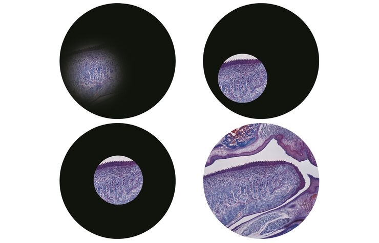
在任何特定光学显微镜调试当中,柯勒照明都是实现最佳成像的最重要且最基本的技术之一。尽管柯勒照明应该视为显微镜设置的常规组成部分,但是许多显微镜学家认为正确的设置过于复杂耗时,因此仍然没有被广泛应用。充分了解显微镜的两个主要部件(光阑和载物台下聚光透镜)在实际操作中的调整后,正确的设置就只需要几分钟的时间。正确对齐的显微镜可以大大改善图像的均匀对比度和照明,从而获得更高的分辨率和观察到更多的细节。在…
Loading...
![[Translate to chinese:] Left: Tissue cells marked with an immunolabel (FITC) illuminated with wide-band UV excitation. Note the tissue structure with blue autofluorescence. Right: Same tissue and same immunostaining with FITC label illuminated with epi-il [Translate to chinese:] Left: Tissue cells marked with an immunolabel (FITC) illuminated with wide-band UV excitation. Note the tissue structure with blue autofluorescence. Right: Same tissue and same immunostaining with FITC label illuminated with epi-illumination using narrow-band blue (490 nm) light. Note the increased image contrast (Ploem, 1967)](/fileadmin/_processed_/c/2/csm_Ploem_Figure_5_Autofluorescence_a_b_3ad909dc27.png)
Milestones in Incident Light Fluorescence Microscopy
Since the middle of the last century, fluorescence microscopy developed into a bio scientific tool with one of the biggest impacts on our understanding of life. Watching cells and proteins with the…
Loading...
![[Translate to chinese:] Center a fluorescence bulb. [Translate to chinese:] Center a fluorescence bulb.](/fileadmin/_processed_/b/1/csm_Center_Fluorescence_Bulb_02_920d77ed68.jpg)
视频教程: 如何调节荧光光源的灯泡位置
荧光激发的传统光源是带汞灯的荧光灯管。实现明亮均匀激发的前提条件是灯泡在灯罩内正确对中和对齐。
本视频教程介绍了一种简易的方法,用于调节荧光灯管中的汞灯的位置。
Loading...

Optical Contrast Methods
Optical contrast methods give the potential to easily examine living and colorless specimens. Different microscopic techniques aim to change phase shifts caused by the interaction of light with the…
Loading...
![[Translate to chinese:] Tartaric acids, polarization [Translate to chinese:] Tartaric acids, polarization](/fileadmin/_processed_/8/5/csm_Tartaric_acids_polarization_01_8cdd315278.jpg)
偏振光显微观察
偏光显微镜通常应用于材料科学和地质学领域,根据矿物的折射特性和颜色来识别矿物。在生物学中,偏光显微镜通常用于晶体等双折射结构的识别或成像,或用于植物细胞壁中纤维素和淀粉粒的成像。
Loading...
![[Translate to chinese:] Transgenic Mouse Embryo, GFP [Translate to chinese:] Transgenic Mouse Embryo, GFP](/fileadmin/_processed_/f/6/csm_Transgenic_Mouse_Embryo_GFP_e5a2d10fb2.jpg)
显微镜中的荧光
荧光显微技术是一种特殊的光学显微镜技术。它利用的是荧光色素在一定波长的光激发下发光的能力。通过抗体染色或荧光蛋白标记,可以用这种荧光色素标记感兴趣的蛋白质。这样就可以确定单分子物种的分布、数量及其在细胞内的定位。此外,还可以进行共定位和相互作用研究,使用可逆结合染料(如 Ca2+ 和 fura-2)观察离子浓度,以及观察细胞的内吞和外吞过程。如今,利用荧光显微镜甚至可以对亚分辨率颗粒进行成像。

![[Translate to chinese:] Exchange a fluorescence bulb. [Translate to chinese:] Exchange a fluorescence bulb.](/fileadmin/_processed_/6/2/csm_Exchange_Fluorescence_Bulb_02_b569d809dd.jpg)
![[Translate to chinese:] Jellyfish Aequorea Victoria [Translate to chinese:] Jellyfish Aequorea Victoria](/fileadmin/_processed_/7/8/csm_Aequorea3_03_5f0d7319f4.jpg)