
Loading...

选择牙科显微镜时需考虑的六大特性
在牙医学中,手术显微镜对于进行高质量和成功的手术来说变得愈发重要,尤其是在牙髓病学领域。显微镜协助牙医进行微创手术,旨在保护牙质、保留组织、最大限度地降低风险并缩短愈合时间。
要选择适合牙医需求的显微镜,了解现代牙科显微镜的一些决定性特征将十分有帮助。
Loading...
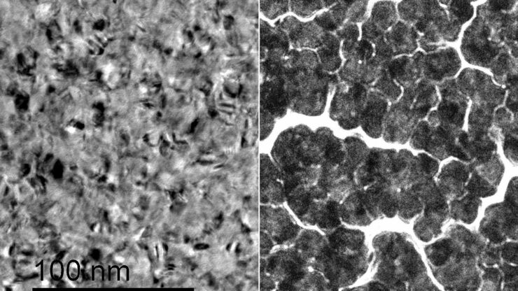
Improvement of Metallic Thin Films for HR-SEM by Using DC Magnetron Sputter Coater
Preparation techniques, like several kinds of coating methods play an important role for high resolution scanning electron microscopy (HR-SEM). Nonconductive sample like biological and synthetic…

Loading...

无限远光学系统
“无限远光学”这一概念是指在显微镜的物镜和镜筒透镜之间具有平行光线的光束路径。平面光学元件可以进入到这个“无限远空间”中,而不影响成像,这对于利用DIC或荧光等对比度方法至关重要。
现代显微技术需要在无限远光路中添加多种光学仪器,如光源或激光装置。满足这一需求的不同方法已经出现,本文对其进行了描述。
Loading...

Imaging of Host Cell-bacteria Interactions using Correlative Microscopy under Cryo-conditions
Pathogenic bacteria have developed intriguing strategies to establish and promote infections in their respective hosts. Most bacterial pathogens initiate infectious diseases by adhering to host cells…
Loading...

Image gallery: Life Science Imaging with the Leica DVM6 Digital Microscope
Digital microscopes can be a great help in life science applications such as the documentation in botany, entomology studies and crop science, or the digitization of museum collections. The image…
Loading...
![[Translate to chinese:] Proveo lightbeam](/fileadmin/_processed_/3/6/csm_proveo_lightbeam_1200x1800_81763a1d26.jpg "[Translate to chinese:] Proveo lightbeam")
使用CoAx 4 四光束同轴立体照明技术实施的白内障手术
稳定的红光反射是白内障手术所用的眼科手术显微镜的最重要功能之一。红光反射让手术医生可以观察到晶状体结构,为其安全成功地实施手术提供清晰的视野。如何能清晰的观察到晶状体结构,特别是在手术过程中的超声乳化、晶状体摘除以及人工晶状体植入等关键阶段,始终提供稳定的红光反射,是手术显微镜面临的挑战。
…
Loading...
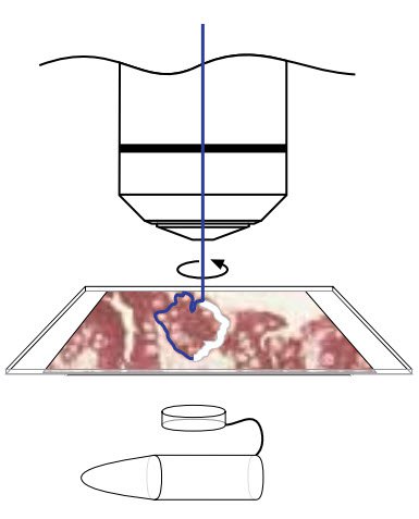
Workflows & Protocols: How to Isolate Individual Chromosomes with Laser Microdissection
During the first Leica Workshop in Brazil, at the Centro de Energia Nuclear na Agricultura/USP (CENA), the participants learned how to prepare samples for laser microdissection (LMD) using a cryotome.…
Loading...
")
A Brief History of Light Microscopy – From the Medieval Reading Stone to Super-Resolution
The history of microscopy begins in the Middle Ages. As far back as the 11th century, plano-convex lenses made of polished beryl were used in the Arab world as reading stones to magnify manuscripts.…