Loading...

A Brief History of Light Microscopy – From the Medieval Reading Stone to Super-Resolution
The history of microscopy begins in the Middle Ages. As far back as the 11th century, plano-convex lenses made of polished beryl were used in the Arab world as reading stones to magnify manuscripts.…
Loading...

From Light to Mind: Sensors and Measuring Techniques in Confocal Microscopy
This article outlines the most important sensors used in confocal microscopy. By confocal microscopy, we mean "True Confocal Scanning", i.e. the technique that illuminates and measures one single…
Loading...

Video: The White Light Laser – How to Effectively Excite Multiple Fluorophores with a Single Light Source
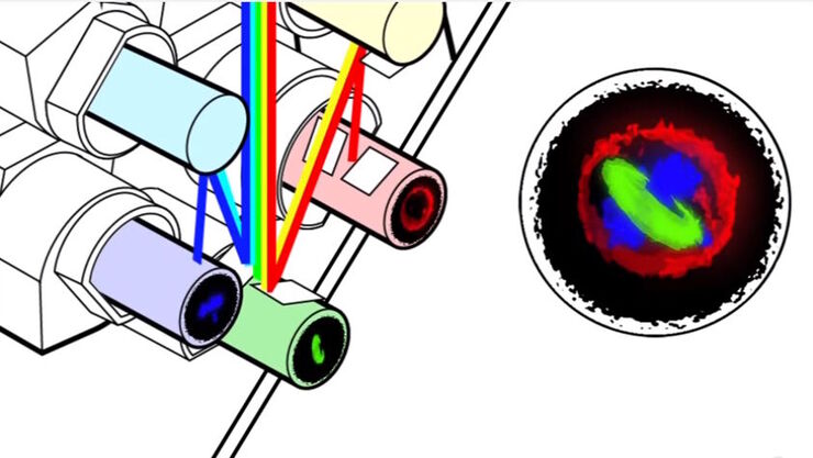
The Leica White Light Laser produces a continuous spectral output between the wavelengths of 470 and 670 nm. It allows you to select 8 excitation lines from 3 trillion unique combinations for…
Loading...

Joseph Gall的视频讲座:显微镜的早期历史
Joseph Gall带我们了解早期显微镜的历史和细胞的发现。复式显微镜是在17世纪与望远镜一起发明的;然而,由于光学畸变,这些显微镜直到19世纪末才被广泛使用。在此期间,简单的显微镜在整个18世纪和19世纪被用于生物学的重大发现,包括对细胞核、纤毛、细胞、细菌和原生动物的首次描述。光学技术在19世纪中后期得到改善厚,复合显微镜就被用来发现染色体、有丝分裂和其他细胞结构。
Loading...

Workflows & Protocols: How to Use a Leica Laser Microdissection System and Qiagen Kits for Successful RNA Analysis
Laser Microdissection (LMD) allows isolating individual cells or chromosomes and is a well established technique for sample preparation prior downstream analysis of the nucleic acid content via PCR or…
Loading...

Confocal and Light Sheet Imaging with STELLARIS DLS
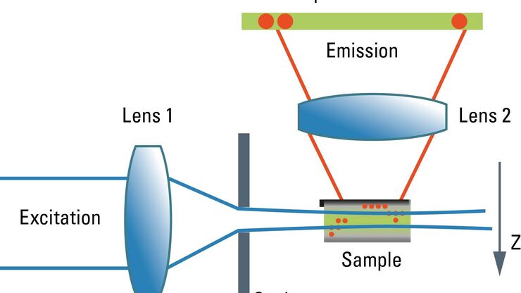
Optical imaging instrumentation can magnify tiny objects, zoom in on distant stars and reveal details that are invisible to the naked eye. But it notoriously suffers from an annoying problem: the…
Loading...

Universal PAINT – Dynamic Super-Resolution Microscopy
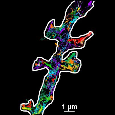
Super-resolution microscopy techniques have revolutionized biology for the last ten years. With their help cellular components can now be visualized at the size of a protein. Nevertheless, imaging…
Loading...

Nanoscale or Microscale Structures Formed in Polymers Containing Nanotubes Greatly Enhance the Electrical Conductivity
The excellent mechanical and electrical properties of carbon nanotubes have led to them being exploited for the creation of a new class of high performance polymer composites. Due to important…
Loading...

冷冻透射电子显微镜的投入式冷冻技术:应用
低温下观察完全含水、未染色样本的透射电子显微镜(cryo TEM)是结构生物学、细胞生物学、药理学和其他科学分支的通用工具。通过将标本放入冷冻剂中进行超快速冷冻(投入式冻结)是一种常用的方法,用于制备在透射电镜观察的各种标本。本文是对投入式冷冻的补充,介绍了在不同领域使用投入式冷冻标本的三种冷冻TEM应用。
