
Loading...

A Meta-cancer Analysis of the Tumor Spatial Microenvironment
Learn how clustering analysis of Cell DIVE datasets in Aivia can be used to understand tissue-specific and pan-cancer mechanisms of cancer progression
Loading...
 tissue.")
Mapping the Landscape of Colorectal Adenocarcinoma with Imaging and AI
Discover deep insights in colon adenocarcinoma and other immuno-oncology realms through the potent combination of multiplexed imaging of Cell DIVE and Aivia AI-based image analysis
Loading...

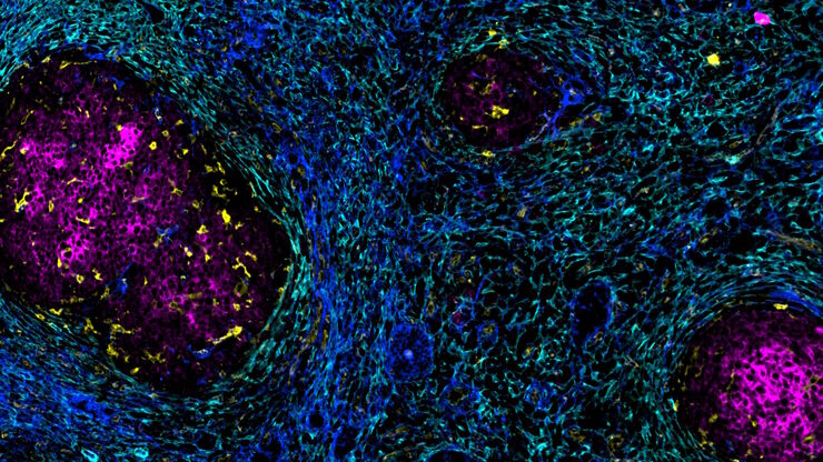
Spatial Architecture of Tumor and Immune Cells in Tumor Tissues
Dig deep into the spatial biology of cancer progression and mouse immune-oncology in this poster, and learn how tumor metabolism can effect immune cell function.
Loading...

IBEX, Cell DIVE, and RNA-Seq: A Multi-omics Approach to Follicular Lymphoma
In a recent study by Radtke et al., a multi-omics spatial biology approach helps shed light on early relapsing lymphoma patients
Loading...
![[Translate to chinese:] Multicolor fixed STED image. Inner ear section, mouse, TauSTED Xtend 589 on AF488 and TauSTED Xtend 775 on AF633-Phalloidin. Sample courtesy of Dennis Derstrof, Klinik für Hals-, Nasen und Ohrenheilkunde, Universität Marburg & Prof. Dr. Dominik Oliver aus dem Institut für Physiologie und Pathophysiologie, Abteilung für Neurophysiologie, Universität Marburg.](/fileadmin/_processed_/c/a/csm_Inner_ear_section_mouse_multicolor_fixed_TauSTED_Xtend_1a1a0b424b.jpg "[Translate to chinese:] Multicolor fixed STED image. Inner ear section, mouse, TauSTED Xtend 589 on AF488 and TauSTED Xtend 775 on AF633-Phalloidin.")
细胞活成像的纳米级扩展
新的STED显微技术方法——TauSTED Xtend,使得在纳米级别下对活体完整样本进行扩展多色成像成为可能。通过结合空间和寿命信息,TauSTED Xtend提供了额外一层信息,允许在极低的光剂量下分辨小细节并在整体结构中解析它们。
Loading...
Accelerating Discovery for Multiplexed Imaging of Diverse Tissues
Explore IBEX: Open-source multiplexed imaging. Join the collaborative IBEX Imaging Community for optimized tissue processing, antibody selection, and human atlas construction.
Loading...

Transforming Multiplexed 2D Data into Spatial Insights Guided by AI
Aivia 13 handles large 2D images and enables researchers to obtain deep insights into microenvironment surrounding their phenotypes with millions of detected objects and automatic clustering up to 30…
![[Translate to chinese:] Hepatocellular Carcinoma with 13 biomarkers shown – Beta-Catenin, CD3D, CD4, CD8a, CD31, CD44, CD163, DAPI, PanCK, PCK26, PD1, SMA, and Vimentin.](/fileadmin/_processed_/5/b/csm_Hepatocellular_Carcinoma_13_Markers_Zoom2_bc27c21cf2.jpg "[Translate to chinese:] Hepatocellular Carcinoma with 13 biomarkers shown – Beta-Catenin, CD3D, CD4, CD8a, CD31, CD44, CD163, DAPI, PanCK, PCK26, PD1, SMA, and Vimentin.")
Loading...
![[Translate to chinese:] Adult human Alzheimer’s brain demonstrating a panel of 15 markers.](/fileadmin/_processed_/4/9/csm_Adult_human_Alzheimers_brain_68bacde06e.jpg "[Translate to chinese:] Adult human Alzheimer’s brain demonstrating a panel of 15 markers.")
大脑的形状:阿尔茨海默病的空间生物学
阿尔茨海默病(AD)是一种神经退行性疾病,也是导致晚年认知障碍的常见原因。阿尔茨海默病的特征是出现含β-淀粉样蛋白的斑块和含磷酸化 tau 的神经纤维缠结。目前尚缺乏治疗和预防AD的有效疗法。我们将Cell DIVE与Cell Signaling Technology的抗体结合使用,检查了AD的突触过程并从空间上确定了神经胶质细胞和神经元等细胞,证明了超多标免疫荧光成像技术可用于探测AD大脑。