
Loading...

超薄切片介绍
对样本开展研究时,为了以纳米级分辨率显示其精细结构,通常会使用到电子显微镜。电子显微镜有两种类型:扫描电子显微镜(SEM)用于对样本表面成像,以及需要使用极薄电子透明样本的透射电子显微镜(TEM)。因此,使用电子显微镜对样本内部的精细结构进行成像时,此类技术解决方案需要制作出非常薄的样本切片。被称为超显微技术的样本制备方法可以产生具有最小伪影的超薄切片(厚度20-150nm)。在切片过程中,样本的…
Loading...
![[Translate to chinese:] Roland A. Fleck](/fileadmin/_processed_/6/1/csm_interview-fleck-teaser_3a7e2a45ad.jpg "[Translate to chinese:] Roland A. Fleck")
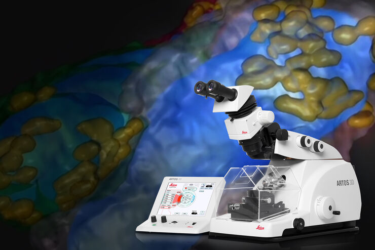
专家在低温扫描电镜工作流程高压冷冻和冷冻断裂方面的知识
深入了解实验室工作方法并了解在EM样本制备过程中低温扫描电镜研究的优势。了解如何将高压冷冻、冷冻断裂和冷冻传送添加到低温扫描电镜工作流程中,以及徕卡组合如何确保这些不同步骤之间的兼容性。
Loading...
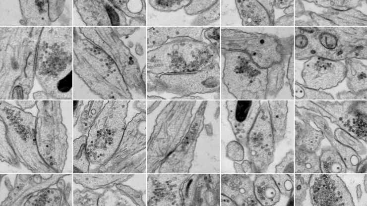
通过光遗传和电刺激技术研究纳米桥接结构和动力学
纳米级超微结构信息通常是由经固定和处理样品的静态图像获得的。但是,这些静态图像只是不断变化的动态结构中的一个瞬间。因此,如何探索动态过程中的特定时间点,是纳米级超微结构研究的一个重大挑战。通过光遗传或电刺激技术,并结合毫秒级样品玻璃化技术探索纳米级超微结构,是一种解决上述问题具有前景的技术。在本应用白皮书的第一部分中,我们将从实际应用角度讨论光刺激辅助的样品玻璃化工作流程。
Loading...
Array Tomography for SEM 3D Reconstruction
Array tomography (AT) is a 3D image reconstruction technique for high resolution, quantitative analysis of biological structures. For optimal results, ultrathin and ordered sections are an absolute…
Loading...
Expanding the Limits of Electron Microscopy Sample Preparation
Capturing the intricate changes in fine structure or in cell dynamics with conventional cryo solutions can be challenging sometimes. Leica Microsystems has developed a new cryo platform, the Leica EM…
![[Translate to chinese:] Array tomography image of T-cells in mouse lymph nodes.](/fileadmin/_processed_/b/9/csm_T-cells_in_mouse_lymph_nodes_array_tomography_d9e5d6f4da.jpg "[Translate to chinese:] Array tomography image of T-cells in mouse lymph nodes.")
Loading...
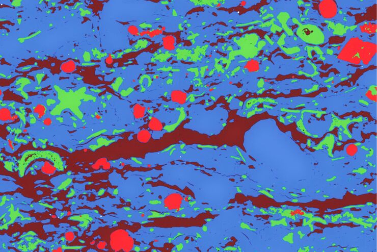
Macroscale to Nanoscale Pore Analysis of Shale and Carbonate Rocks
Physical porosity in rocks, like shale and carbonate, has a large effect on the their storage capacity. The pore geometries also affect their permeability. Imaging the visible pore space provides…
Loading...
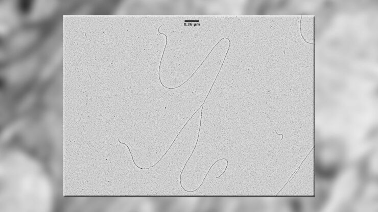
Visualization of DNA Molecules
Precise low angle rotary shadowing with heavy metals (like platinum) can be used in transmission electron microscopy (TEM) to observe molecular details of objects previously absorbed on a thin, low…
Loading...
Porous Ceramics - Sample Preparation for SEM
Application Note for Leica EM RES102 - Ceramic membrane filters with pore sizes down to a few nanometres must be investigated in cross-section with regard to the structure of the pores. The smallest…