Loading...
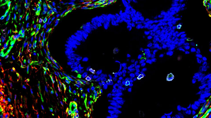
A Meta-cancer Analysis of the Tumor Spatial Microenvironment
Learn how clustering analysis of Cell DIVE datasets in Aivia can be used to understand tissue-specific and pan-cancer mechanisms of cancer progression
Loading...
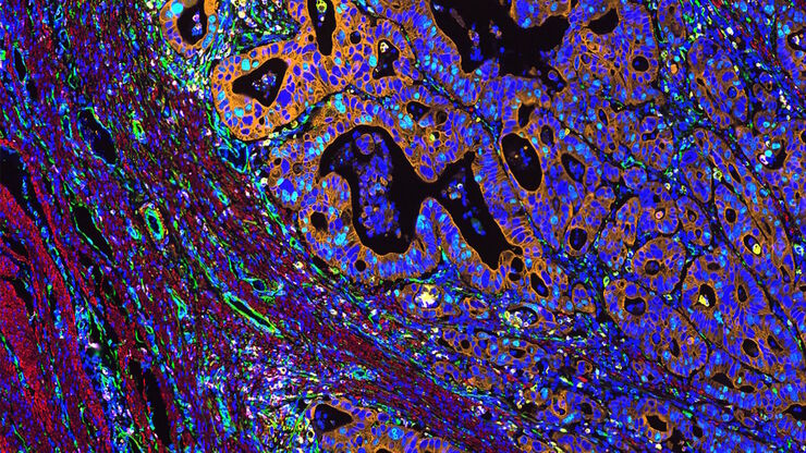
Mapping the Landscape of Colorectal Adenocarcinoma with Imaging and AI
Discover deep insights in colon adenocarcinoma and other immuno-oncology realms through the potent combination of multiplexed imaging of Cell DIVE and Aivia AI-based image analysis
Loading...
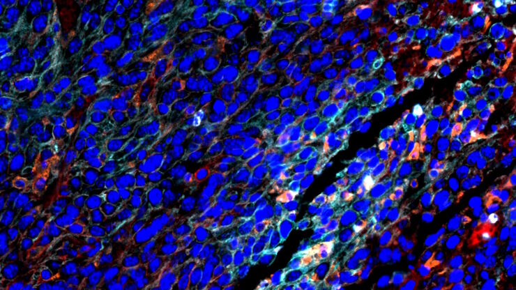
Spatial Architecture of Tumor and Immune Cells in Tumor Tissues
Dig deep into the spatial biology of cancer progression and mouse immune-oncology in this poster, and learn how tumor metabolism can effect immune cell function.
Loading...
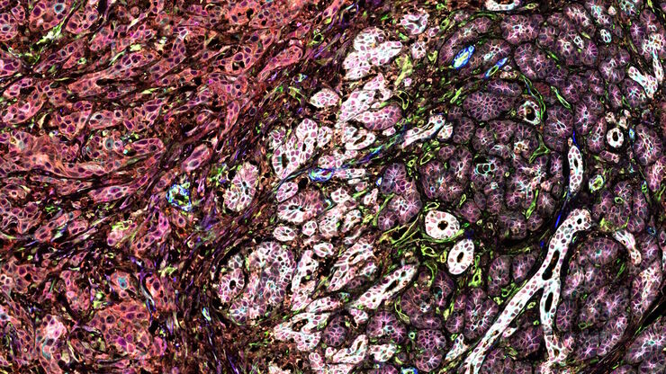
IBEX, Cell DIVE, and RNA-Seq: A Multi-omics Approach to Follicular Lymphoma
In a recent study by Radtke et al., a multi-omics spatial biology approach helps shed light on early relapsing lymphoma patients
Loading...
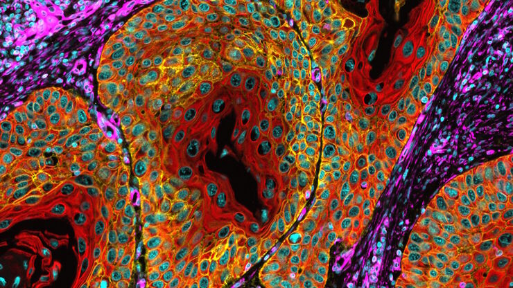
Discover how Multiplexed Bioimaging can Advance Cancer Research
Explore multiplexing with up to 60 biomarkers, enabling advanced tumor imaging approaches to gather precise, spatially-resolved single-cell data that helps enhance cancer research and clinical…
Loading...
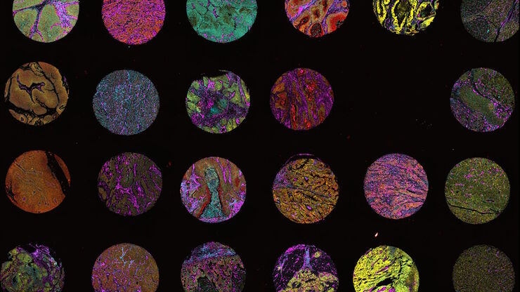
Spatial Biology: Learning the Landscape
Spatial Biology: Understanding the organization and interaction of molecules, cells, and tissues in their native spatial context
Loading...
![[Translate to chinese:] Branched organoid growing in collagen where the Nuclei are labeled blue. To detect the mechanosignaling process, the YAP1 is labeled green. [Translate to chinese:] Branched organoid growing in collagen where the Nuclei are labeled blue. To detect the mechanosignaling process, the YAP1 is labeled green.](/fileadmin/_processed_/a/e/csm_Branched_organoid_growing_in_collagen_083254eb44.jpg)
检查癌症类器官的发展进程
德国慕尼黑工业大学的Andreas Bausch实验室研究细胞和生物体中不同结构和功能形成的细胞和生物物理机制。他的团队设计了新的策略、方法和分析工具,以量化微米和纳米等级的发展机制和动态过程。关键研究领域包括干细胞和类器官,从乳腺类器官到胰腺癌类器官,以更好地了解疾病模型。

![[Translate to chinese:] Spheroid stained with Cyan: Dapi nuclear countertain; Green AF488 Involucrin; Orange AF55 Phalloidin Actin; Magenta AF647 CK14. [Translate to chinese:] Spheroid stained with Cyan: Dapi nuclear countertain; Green AF488 Involucrin; Orange AF55 Phalloidin Actin; Magenta AF647 CK14.](/fileadmin/_processed_/6/8/csm_Spheroid_stained_with_Cyan_4color_overlay_76cb6e4d04.jpg)
![[Translate to chinese:] Hepatocellular Carcinoma with 13 biomarkers shown – Beta-Catenin, CD3D, CD4, CD8a, CD31, CD44, CD163, DAPI, PanCK, PCK26, PD1, SMA, and Vimentin. [Translate to chinese:] Hepatocellular Carcinoma with 13 biomarkers shown – Beta-Catenin, CD3D, CD4, CD8a, CD31, CD44, CD163, DAPI, PanCK, PCK26, PD1, SMA, and Vimentin.](/fileadmin/_processed_/5/b/csm_Hepatocellular_Carcinoma_13_Markers_Zoom2_bc27c21cf2.jpg)