
Loading...

超薄切片介绍
对样本开展研究时,为了以纳米级分辨率显示其精细结构,通常会使用到电子显微镜。电子显微镜有两种类型:扫描电子显微镜(SEM)用于对样本表面成像,以及需要使用极薄电子透明样本的透射电子显微镜(TEM)。因此,使用电子显微镜对样本内部的精细结构进行成像时,此类技术解决方案需要制作出非常薄的样本切片。被称为超显微技术的样本制备方法可以产生具有最小伪影的超薄切片(厚度20-150nm)。在切片过程中,样本的…
Loading...
改善冷冻电子断层扫描工作流程
徕卡显微系统有限公司和赛默飞世尔科技有限公司合作开发了一个整条技术路线的冷冻电子断层扫描工作流程。它确保从通过THUNDER成像仪EM冷冻CLEM(也可选择新版的CORAL Cryo冷冻共聚焦CLEM)预选与我们的EM GP2的玻璃化冷冻到Thermo Scientific Krios™ G3i Cryo TEM的3D图像重建的完全整合。所有仪器之间的无缝通信能够获得可靠的结果和可重现的实验。
Loading...
![3D glomeruli in a portion of an ECi-cleared kidney scanned by light sheet microscopy. Courtesy of Prof. Norbert Gretz, Medical Faculty Mannheim, University of Heidelberg [1].](/fileadmin/_processed_/d/d/csm_DLS-Sample-Preparation-Intr_527af241c5.jpg "3D glomeruli in a portion of an ECi-cleared kidney scanned by light sheet microscopy. Courtesy of Prof. Norbert Gretz, Medical Faculty Mannheim, University of Heidelberg [1].")
Using Mounting Frames for Light Sheet Sample Preparation
Sample handling is an important topic in the context of Light Sheet Microscopy. The TCS SP8 DLS integrates Light Sheet technology into an inverted confocal platform and can hence make use of general…
Loading...

Using a Rotation Device for Light Sheet Sample Mounting
The TCS SP8 DLS from Leica Microsystems is an innovative concept to integrate the Light Sheet Microscopy technology into the confocal microscope. Due to its unique optical architecture samples can be…
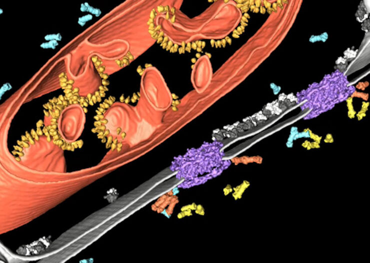
![[Translate to chinese:] Array tomography image of T-cells in mouse lymph nodes.](/fileadmin/_processed_/b/9/csm_T-cells_in_mouse_lymph_nodes_array_tomography_d9e5d6f4da.jpg "[Translate to chinese:] Array tomography image of T-cells in mouse lymph nodes.")
Loading...
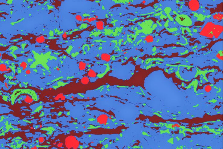
Macroscale to Nanoscale Pore Analysis of Shale and Carbonate Rocks
Physical porosity in rocks, like shale and carbonate, has a large effect on the their storage capacity. The pore geometries also affect their permeability. Imaging the visible pore space provides…
Loading...

Using U-Shaped Glass Capillaries for Sample Mounting
The DLS microscope system from Leica Microsystems is an innovative concept which integrates the Light Sheet Microscopy technology into the confocal platform. Due to its unique optical architecture,…
Loading...
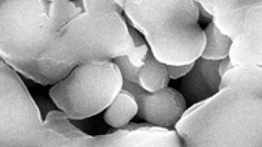
Porous Ceramics - Sample Preparation for SEM
Application Note for Leica EM RES102 - Ceramic membrane filters with pore sizes down to a few nanometres must be investigated in cross-section with regard to the structure of the pores. The smallest…
Loading...
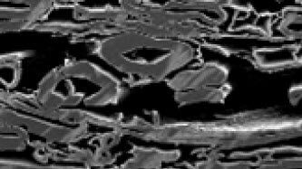
Paper Samples - Sample Preparation for SEM
Application Note for Leica EM RES102 - A coated paper sample has been prepared with ion beam slope cutting in order to test the procedure with regard to its applicability. With the use of ion beam…