
Loading...
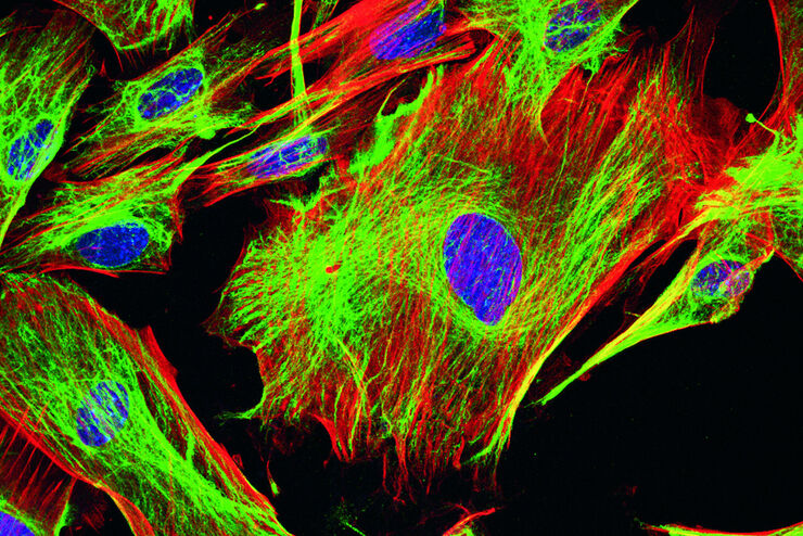
荧光染料
荧光显微镜的基本原理是借助荧光染料对细胞成分进行高度特异性的可视化观察。这可能是一种与兴趣蛋白质遗传相关的荧光蛋白,如绿色荧光蛋白(GFP)等。如果克隆无法实现,例如在组织学样本上无法实现,则需要使用另一种技术如免疫荧光染色来对兴趣蛋白质进行可视化观察。为此,人们使用抗体来连接不同的荧光染料并将其直接或间接地结合到适当的靶点上。此外,借助荧光染料,荧光显微镜的应用就不再仅局限于蛋白质观察,还能对核…
Loading...
利用多重中频成像设计您的研究课题
多重组织分析是一种功能强大的技术,可对单个固定组织样本中的细胞类型位置和细胞类型相互作用进行比较。在多重分析研究开始之前,研究人员通常会提出以下问题: "我如何知道组织中哪些生物标记物是相关的?另外,随着研究问题的发展,我如何转向其他生物标记物?巧妙的研究设计有助于回答现有的问题,并能继续探索研究开始时并不明显的新联系。
Loading...
 - THUNDER Imager 3D Cell Culture Influenca virus – red, cilia – green, Nuclei – blue.")
How Can Immunofluorescence Aid Virology Research?
Modern virology research has become as crucial now as ever before due to the global COVID-19 pandemic. There are many powerful technologies and assays that virologists can apply to their research into…
Loading...

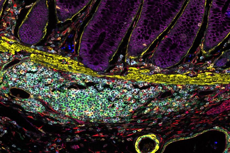
Chronic Inflammation Under the Microscope
In the course of chronic inflammation certain body areas are recurrently inflamed. This goes along with many human diseases. With the help of widefield light microscopy, the underlying processes can…
Loading...
BABB Clearing and Imaging for High Resolution Confocal Microscopy
Multipohoton microscopy experiment using Leica TCS SP8 MP and Leica 20x/0.95 NA BABB immersion objective.
Understanding kidney microanatomy is key to detecting and identifying early events in kidney…
Loading...
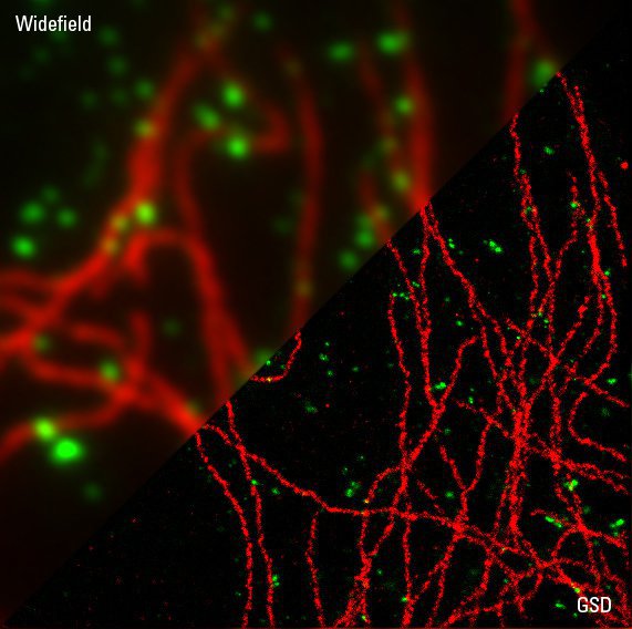
Sample Preparation for GSDIM Localization Microscopy – Protocols and Tips
The widefield super-resolution technique GSDIM (Ground State Depletion followed by individual molecule return) is a localization microscopy technique that is capable of resolving details as small as…